The nervous system plays an exceptional integrating role in the life of the organism, since it unites (integrates) it into a single whole and "inscribes" (integrates) it into the environment. It ensures the coordinated work of individual parts of the body ( coordination), maintaining an equilibrium state in the body ( homeostasis) and adaptation of the body to changes in the external and / or internal environment ( adaptive state and / or adaptive behavior).
The most important thing is that the nervous system does
The nervous system provides the relationship and interaction between the body and the external environment. And for this it does not need so many processes.
Basic processes in the nervous system
1. Transduction ... The transformation of irritation external to the nervous system itself into nervous excitement with which it can operate.
2. Transformation ... Alteration, transformation of the input stream of excitation into an output stream with different characteristics.
3. Distribution ... Distribution of excitation and its direction along different paths, to different addresses.
4. Modeling. Construction of a nervous model of irritation and / or stimulus, which replaces the stimulus itself. The nervous system can work with this model, it can store it, modify it and use it instead of a real stimulus. Sensory image is one of the variants of neural stimulation models.
5. Modulation ... The nervous system, under the influence of irritation, changes itself and / or its activity.
Modulation types
1. Activation (excitement). An increase in the activity of the nervous structure, an increase in its excitation and / or excitability. Dominant state.
2. Oppression (inhibition, inhibition). Decrease in the activity of the nervous structure, inhibition.
3. Plastic reconstruction of the nervous structure.
Plastic remodeling options:
1) Sensitization - improving the transmission of arousal.
2) Habituation - impairment of the transmission of excitement.
3) Temporary neural connection - the creation of a new way of transmitting excitement.
6. Activation of the executive body to take an action. In this way, the nervous system provides reflex response to irritation .
© 2012-2017 Sazonov V.F. © 2012-2016 kineziolog.bodhy.ru ..
Tasks and activities of the nervous system
1. Produce reception - to catch a change in the external environment or internal environment of the body in the form of irritation (this is carried out by sensory systems with the help of their sensory receptors).
2. Produce transduction - transformation (coding) of this irritation into nervous excitement, i.e. a stream of nerve impulses with special characteristics corresponding to irritation.
3. Implement holding - to deliver excitement along the nerve pathways to the necessary parts of the nervous system and to the executive organs (effectors).
4. Produce perception - to create a nervous model of irritation, i.e. build his sensory image.
5. Produce transformation - to convert sensory excitation into effector for the implementation of a response to a change in the environment.
6. Rate results its activities through feedbacks and reverse afferentation.
The importance of the nervous system:
1. Provides interconnection between organs, organ systems and between individual parts of the body. It is her coordination function. It coordinates (coordinates) the work of individual bodies into a single system.
2. Provides interaction of the body with the environment.
3. Provides thought processes. This includes the perception of information, assimilation of information, analysis, synthesis, comparison with past experience, the formation of motivation, planning, goal setting, correction of action when the goal is achieved (correction of errors), performance evaluation, information processing, formation of judgments, conclusions, conclusions and abstract (general) concepts.
4. Carries out control over the state of the organism and its individual parts.
5. Manages the work of the body and its systems.
6. Provides activation and maintenance of tone, i. E. the working state of organs and systems.
7. Supports the vital functions of organs and systems. In addition to the signaling function, the nervous system also has a trophic function, i.e. the biologically active substances released to it contribute to the vital activity of the innervated organs. Organs deprived of such a "nourishment" from nerve cells atrophy; grow wither and may die off.
The structure of the nervous system
Rice.General structure of the nervous system (diagram).© 2017 Sazonov V.F ..

Rice. Scheme of the structure of the central nervous system (central nervous system). A source: Atlas of Physiology. In two volumes. Volume 1: textbook. allowance / A.G. Kamkin, I.S.Kiseleva - 2010 .-- 408 p. (http: //vmede.org/sait/? page = 7 & id = Fiziologiya_atlas_kamakin_2010 & menu = Fiz ...)
Video: central nervous system
The nervous system is functionally and structurally divided into peripheral and central nervous system (CNS).
The central nervous system consists of head and dorsal brain.
The brain is located inside the cerebral section of the skull, and the spinal cord is located in the vertebral canal.
The peripheral part of the nervous system consists of nerves, i.e. bundles of nerve fibers that extend beyond the brain and spinal cord and are sent to various organs of the body. It also includes nerve nodes, or ganglia- clusters of nerve cells outside the spinal cord and brain.
The nervous system functions as a whole.
Nervous system functions:
1) the formation of excitement;
2) transfer of excitement;
3) inhibition (cessation of arousal, a decrease in its intensity, oppression, limitation of the spread of excitement);
4) integration (combining various streams of excitation and changes in these streams);
5) the perception of irritation from the external and internal environment of the body with the help of special nerve cells - receptors;
6) coding, i.e. transformation of chemical, physical irritation into nerve impulses;
7) trophic, or nutritional, function - the formation of biologically active substances (BAS).
Neuron

Definition of the concept
The neuron is the basic structural and functional unit of the nervous system.
Neuron
is a specialized dendritic cell capable of perceiving, conducting and transmitting nervous excitement for processing information in the nervous system. © 2016 Sazonov V.F ..
A neuron is a complex structure excitable secreting highly differentiated nerve cell with shoots, which perceives nervous excitement, processes it and transfers it to other cells. In addition to the excitatory effect, a neuron can also exert an inhibitory or modulating effect on its target cells.
The work of the inhibitory synapse
The inhibitory synapse has receptors on its postsynaptic membrane to the inhibitory mediator - gamma-aminobutyric acid (GABA or GABA). In contrast to the excitatory synapse in the inhibitory synapse on the postsynaptic membrane, GABA opens ion channels not for sodium, but for chlorine. Chlorine ions do not bring a positive charge into the cell, but a negative one, therefore they counteract excitation, because neutralize the positive charges of sodium ions that excite the cell.
Video:The work of the GABA receptor and inhibitory synapse
So, excitation through synapses is transmitted chemically with the help of special control substances,located in synaptic vesicles located in the presynaptic plaque... The common name for these substances is neurotransmitters
, i.e. "neurotransmitters". They are divided intomediators (intermediaries) that transmit excitation or inhibition, and modulators, which change the state of the postsynaptic neuron, but do not transmit excitation or inhibition themselves.
Nervous system consists of tortuous networks of nerve cells that make up various interconnected structures and control all the body's activities, both desired and conscious actions, as well as reflexes and automatic actions; the nervous system allows us to interact with the outside world, and is also responsible for mental activity.
The nervous system consists from various interconnected structures that together constitute an anatomical and physiological unit. consists of organs located inside the skull (brain, cerebellum, brain stem) and the spine (spinal cord); is responsible for interpreting the state and various needs of the body based on the information received, in order to then generate commands designed to obtain reasonable answers.
consists of many nerves that go to the brain (brain pairs) and the spinal cord (vertebral nerves); acts as a transmitter of sensory stimuli to the brain and commands from the brain to the organs responsible for their execution. The autonomic nervous system controls the functions of numerous organs and tissues through antagonistic effects: the sympathetic system is activated during anxiety, and the parasympathetic system at rest.

central nervous system Includes the spinal cord and brain structures.
The entire nervous system is divided into central and peripheral. The central nervous system includes the brain and spinal cord. From them, nerve fibers spread throughout the body - the peripheral nervous system. It connects the brain with the senses and with the executive organs - muscles and glands.
All living organisms have the ability to respond to physical and chemical changes in their environment.
The stimuli of the external environment (light, sound, smell, touch, etc.) are converted by special sensitive cells (receptors) into nerve impulses - a series of electrical and chemical changes in the nerve fiber. Nerve impulses are transmitted along sensory (afferent) nerve fibers to the spinal cord and brain. Here, the corresponding command impulses are generated, which are transmitted along motor (efferent) nerve fibers to the executive organs (muscles, glands). These executive organs are called effectors.
The main function of the nervous system is the integration of external influences with the corresponding adaptive response of the body.
The structural unit of the nervous system is a nerve cell - neuron. It consists of a cell body, a nucleus, branched processes - dendrites - along them, nerve impulses go to the cell body - and one long process - axon - along it, a nerve impulse passes from the cell body to other cells or effectors.
The processes of two neighboring neurons are connected by a special formation - a synapse. It plays an essential role in filtering nerve impulses: it passes some impulses and delays others. Neurons are connected to each other and carry out joint activities.
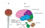
The central nervous system consists of the brain and spinal cord. The brain is subdivided into the brainstem and forebrain. The brain stem consists of the medulla oblongata and the midbrain. The forebrain is subdivided into diencephalon and terminal.
All parts of the brain have their own functions.
So, the diencephalon consists of the hypothalamus, the center of emotions and vital needs (hunger, thirst, libido), the limbic system (in charge of emotional-impulsive behavior) and the thalamus (performing filtering and primary processing of sensory information).
In humans, the cerebral cortex is especially developed - an organ of higher mental functions. It has a thickness of 3 mm, and its total area is on average 0.25 sq. M.
The bark has six layers. The cells of the cerebral cortex are interconnected.
There are about 15 billion of them.
Various neurons in the cortex have their own specific function. One group of neurons performs the function of analysis (cleavage, dismemberment of a nerve impulse), another group carries out synthesis, combines impulses from various sensory organs and parts of the brain (associative neurons). There is a system of neurons that retains traces from previous influences and compares new influences with existing traces.
According to the features of the microscopic structure, the entire cerebral cortex is divided into several tens of structural units - fields, and according to the location of its parts - into four lobes: occipital, temporal, parietal and frontal.
The human cerebral cortex is an integral working organ, although its individual parts (areas) are functionally specialized (for example, the occipital cortex performs complex visual functions, frontal-temporal-speech, temporal-auditory). The largest part of the motor area of the human cerebral cortex is associated with the regulation of the movement of the labor organ (hand) and speech organs.
All parts of the cerebral cortex are interconnected; they are also connected with the underlying parts of the brain, which carry out the most important vital functions. The subcortical formations, regulating innate unconditioned reflex activity, are the area of those processes that are subjectively felt in the form of emotions (they, in the words of I.P. Pavlov, are “a source of strength for cortical cells”).
The human brain contains all those structures that arose at various stages of the evolution of living organisms. They contain the "experience" accumulated in the process of the entire evolutionary development. This testifies to the common origin of humans and animals.
As the organization of animals becomes more complex at different stages of evolution, the importance of the cerebral cortex increases more and more.
If, for example, the cerebral cortex of a frog is removed (it has an insignificant proportion in the total volume of its brain), then the frog hardly changes its behavior. Deprived of the cerebral cortex, the pigeon flies, maintains balance, but already loses a number of vital functions. A dog with a removed cerebral cortex becomes completely unadapted to the environment.
The main mechanism of nervous activity is the reflex. Reflex
The body's response to external or internal influences through the central nervous system.
The term "reflex", as already noted, was introduced into physiology by the French scientist René Descartes in the 17th century. But to explain mental activity, it was applied only in 1863 by the founder of Russian materialistic physiology, M.I. Sechenov. Developing the teachings of I.M.Sechenov, I.P. Pavlov experimentally investigated the features of the functioning of the reflex.
All reflexes are divided into two groups: conditioned and unconditioned.
Unconditioned reflexes are innate reactions of the body to vital stimuli (food, danger, etc.). They do not require any conditions for their production (for example, the blinking reflex, salivation at the sight of food).
Unconditioned reflexes are a natural reserve of ready, stereotyped reactions of the body. They arose as a result of a long evolutionary development of this animal species. Unconditioned reflexes are the same in all individuals of the same species; it is the physiological mechanism of instincts. But the behavior of higher animals and humans is characterized not only by congenital, i.e. unconditioned reactions, but also such reactions that are acquired by a given organism in the process of its individual vital activity, i.e. conditioned reflexes.
Conditioned reflexes are the physiological mechanism of the organism's adaptation to changing environmental conditions.
Conditioned reflexes are those reactions of the body that are not congenital, but are developed in various life conditions.
They arise under the condition of the constant precedence of various phenomena with those that are vital for the animal. If the connection between these phenomena disappears, then the conditioned reflex fades away (for example, the roar of a tiger in a zoo, without being accompanied by its attack, ceases to frighten other animals).
The brain does not go on about only current influences. He plans, anticipates the future, carries out anticipatory reflection of the future. This is the most important feature of his work. The action must achieve a certain future result - the goal. Without the brain's preliminary modeling of this result, regulation of behavior is impossible.
Modern brain science - neurophysiology - is based on the concept of functional unification of brain mechanisms for the implementation of behavioral acts. This concept was put forward and fruitfully developed by the student of I.P. Pavlov, academician P.K. Anokhin in his theory of functional systems.
The functional system P.K. Anokhin calls the unity of central and peripheral neurophysiological mechanisms, which together provide the effectiveness of the behavioral act.
The initial stage of the formation of any behavioral act is called by PK Anokhin afferent synthesis (translated from Latin - “bringing together”).
In the process of afferent synthesis, a variety of information is processed from the external and internal world, based on the currently dominant motivation (need). From the numerous formations of the brain, everything that was associated in the past with the satisfaction of this need is extracted.
Establishing that a given need can be satisfied by a certain action, the choice of this action is called making a decision.
The neurophysiological mechanism of decision-making was named by P.K. Anokhin as an acceptor of the results of action. An acceptor (“assertare” -permissive) of the results of an action is a neurophysiological mechanism for anticipating the results of a future action. Based on the comparison of previously obtained results, an action program is created. And only after that the action itself takes place. The course of action, the effectiveness of its stages, the compliance of these results with the formed action program is constantly monitored by receiving signals about the achievement of the goal. This mechanism of constant receipt of information about the results of the performed action was named by P.K. Anokhin as reverse afferentation.
❖ The human nervous system is represented by:
■ the brain and spinal cord (together they form central nervous system
);
■ nerves, ganglia and nerve endings (form peripheral nervous system
).
Functions of the human nervous system:
■ unites all parts of the body into a single whole ( integration );
■ regulates and harmonizes the work of different organs and systems ( reconciliation );
■ carries out the connection of the organism with the external environment, its adaptation to environmental conditions and survival in these conditions ( reflection and adaptation );
■ provides (in interaction with the endocrine system) the constancy of the internal environment of the body at a relatively stable level ( correction );
■ determines the consciousness, thinking and speech of a person, his purposeful behavioral, mental and creative activity ( activity ).
❖ Division of the nervous system according to functional characteristics:
■ somatic (innervates the skin and muscles; perceives the effects of the external environment and causes contractions of skeletal muscles); obeys the will of man;
■ autonomous , or vegetative (regulates metabolic processes, growth and reproduction, the work of the heart and blood vessels, internal organs and endocrine glands).
Spinal cord
Spinal cord is located in the spinal canal of the spine, starts from the medulla oblongata (above) and ends at the level of the second lumbar vertebra. It is a white cylindrical cord (cord) with a diameter of about 1 cm and a length of 42-45 cm. In front and behind the spinal cord has two deep grooves dividing it into the right and left halves.
In the longitudinal direction of the spinal cord, one can distinguish 31 segments , each with two front and two rear spine formed by the axons of neurons; in this case, all segments form a single whole.
Inside spinal cord is Gray matter , which has (in section) the characteristic shape of a flying butterfly, the "wings" of which form front, rear and (in the thoracic region) side horns .
Gray matter consists of bodies of intercalary and motor neurons. Along the axis of the gray matter along the spinal cord, a narrow spinal drip filled with cerebrospinal fluid (see below).
On the periphery the spinal cord (around the gray matter) is white matter .
White matter located in the form of 6 pillars around the gray matter (two front, side and rear).
It is formed by axons collected in ascending (located in the posterior and lateral columns; transmit excitation to the brain) and downstream (located in the front and side pillars; transmit excitation from the brain to the working organs) pathways spinal cord.
Spinal cord protected by thundering casings: hard (from the connective tissue lining the spinal canal), cobweb (in the form of a thin network; contains nerves and blood vessels) and soft , or vascular (contains many vessels; grows together with the surface of the brain). The space between the arachnoid and soft membranes is filled with cerebrospinal fluid, which provides optimal conditions for the vital activity of nerve cells and protects the spinal cord from shocks and concussions.
V front horns segments of the spinal cord (they are located closer to the abdominal surface of the body) are the body motor neurons , from which their axons depart, forming the anterior motor roots , through which excitation is transmitted from the brain to the working organ (these are the longest human cells, their length can reach 1.3 m).
V rear horns segments are bodies interneurons ; the rear fit them sensitive roots formed by the axons of sensory neurons that transmit excitation to the spinal cord. The bodies of these neurons are in spinal nodes (ganglia) located outside the spinal cord along the sensory neurons.
In the thoracic region there are side horns where the bodies of neurons are located sympathetic parts autonomous nervous system.
Outside the spinal canal, the sensory and motor roots, extending from the posterior and anterior horns of one "wing" of the segment, unite, forming (together with the nerve fibers of the autonomic nervous system) a mixed spinal nerve , which contains both centripetal (sensitive) and centrifugal (motor) fibers (see below).
❖ Functions of the spinal cord carried out under the control of the brain.
■ Reflex function:
pass through the gray matter of the spinal cord arcs of unconditioned reflexes
(they do not affect the consciousness of a person), regulating
the work of internal organs, the lumen of blood vessels, urination, sexual function, contraction of the diaphragm, defecation, sweating, and managers
skeletal muscles; (examples, knee reflex:
lifting the leg when hitting the tendon attached to the patella; limb withdrawal reflex: under the action of a painful stimulus, reflex muscle contraction and withdrawal of the limb occur; reflex urination: filling the bladder triggers the stretch receptors in the bladder wall, which leads to relaxation of the sphincter, contraction of the bladder wall and urination). 
When the spinal cord breaks above the arc of the unconditioned reflex, this reflex does not experience the regulatory action of the brain and is perverted (deviates from the norm, i.e., becomes pathological).
■ Conductive function; the pathways of the white matter of the spinal cord are the conductors of nerve impulses: ascending pathways, nerve impulses from the gray matter of the spinal cord go to the brain (nerve impulses coming from sensory neurons first enter the gray matter of certain segments of the spinal cord, where they undergo preliminary processing), and downward the ways they go from the brain into different segments of the spinal cord and from there along the spinal nerves to the organs.
In humans, the spinal cord controls only simple motor acts; complex movements (walking, writing, work skills) are carried out with the obligatory participation of the brain.
Paralysis- loss of the ability to voluntary movements of the body organs, which occurs when the cervical spinal cord is damaged, resulting in a violation of the connection between the brain and the organs of the body located below the site of injury.
Spinal shock- This is the disappearance of all reflexes and voluntary movements of body organs, the nerve centers of which lie below the site of injury, which occurs when the spine is injured and the connection between the brain and the underlying (in relation to the injury site) parts of the spinal cord is disrupted.
Nerves. Nerve impulse propagation
Nerves- These are cords of nerve tissue that connect the brain and nerve nodes with other organs and tissues of the body through nerve impulses transmitted through them.
Nerves are formed from multiple bundles nerve fibers (up to 106 fibers in total) and a small number of thin blood vessels enclosed in a common connective tissue sheath. For each nerve fiber, the nerve impulse propagates in isolation, without passing to other fibers.
■ Most of the nerves mixed ; they include fibers of both sensory and motor neurons.
Nerve fiber- a long (may have a length of more than 1 m) thin process of a nerve cell ( axon), strongly branching at the very end; serves to transmit nerve impulses.
❖ Classification of nerve fibers depending on the structure: myelinated and unmyelinated .
Myelinated nerve fibers are covered with a myelin sheath. Myelin sheath performs the functions of protection, nutrition and isolation of nerve fibers. It has a protein-lipid nature and is a plasmalemma Schwann cell (named after its discoverer T. Schwann, 1810-1882), which repeatedly (up to 100 times) turns around the axon; at the same time, the cytoplasm, all organelles and the membrane of the Schwann cell are concentrated on the periphery of the membrane above the last turn of the plasmalemma. There are open sections of the axon between adjacent Schwann cells - Ranvier's interceptions ... A nerve impulse along such a fiber spreads in jumps from one interception to another at a high speed - up to 120 m / s.
Unmyelinated the nerve fibers are covered only with a thin insulating sheath that does not contain myelin. The speed of propagation of a nerve impulse along an unmyelinated nerve fiber is 0.2-2 m / s.
Nerve impulse Is a wave of excitation that propagates along the nerve fiber in response to irritation of the nerve cell.
■ The speed of propagation of a nerve impulse along the fiber is directly proportional to the square root of the fiber diameter.
The mechanism of propagation of a nerve impulse. Simplified, a nerve fiber (axon) can be represented as a long cylindrical tube with a surface membrane separating two aqueous solutions of different chemical composition and concentration. The membrane has numerous valves that close when the electric field increases (i.e., when its potential difference increases) and open when it weakens. When open, some of these valves allow Na + ions to pass through, other valves allow K + ions to pass through, but all of them do not allow large ions of organic molecules to pass through.
Each axon is a microscopic powerhouse, separating (through chemical reactions) electrical charges. When the axon not excited , inside it there is an excess (in comparison with the environment surrounding the axon) of potassium cations (K +), as well as negative ions (anions) of a number of organic molecules. Outside the axon, there are sodium cations (Na +) and chlorine anions (C1 -), formed as a result of the dissociation of NaCl molecules. Anions of organic molecules are concentrated on internal membrane surface, charging it negatively , and sodium cations - on its external surface, charging it positively ... As a result, an electric field arises between the inner and outer surfaces of the membrane, the potential difference (0.05 V) of which ( rest potential) is large enough to close the diaphragm valves. Resting potential was first described and measured in 1848-1851. German physiologist E.G. Dubois-Reymond in experiments on the muscles of a frog.
When the axon is irritated, the density of electric charges on its surface decreases, the electric field weakens and the membrane valves open slightly, allowing the sodium cation Na + into the axon. These cations partially compensate for the negative electric charge of the inner surface of the membrane, as a result of which, at the site of stimulation, the direction of the field is reversed. The process involves neighboring sections of the membrane, which gives rise to the propagation of a nerve impulse. At this moment, the valves open, allowing the potassium cations K + to pass outside, due to which a negative charge is gradually restored inside the axon, and the potential difference between the inner and outer surfaces of the membrane reaches a value of 0.05 V, characteristic of an unexcited axon. Thus, it is actually not an electric current that propagates along the axon, but a wave of an electrochemical reaction.
■ The shape and speed of propagation of a nerve impulse does not depend on the degree of irritation of the nerve fiber. If it is very strong, a whole series of identical impulses arises; if it is very weak, the impulse does not appear at all. Those. exists some minimum "threshold" degree of irritation, below which the impulse is not excited.
The impulses entering the neuron along the nerve fiber from any receptor differ only in the number of signals in the series. This means that it is enough for a neuron to count the number of such signals in one series and, in accordance with the "rules", how to react to a given number of sequential signals, to send the necessary command to one or another organ.
Spinal nerves
Each spinal nerve formed from two roots extending from the spinal cord: front (efferent) root and rear (afferent) roots, which connect in the intervertebral foramen, forming mixed nerves (contain motor, sensory and sympathetic nerve fibers).
■ A person has 31 pairs of spinal nerves (according to the number of spinal cord segments) extending to the right and left of each segment.
Spinal nerve functions:
■ they determine the sensitivity of the skin of the upper and lower extremities, chest, abdomen;
■ carry out the transmission of nerve impulses that provide movement of all parts of the body and limbs;
■ innervate skeletal muscles (diaphragm, intercostal muscles, muscles of the walls of the chest and abdominal cavities), causing their involuntary movements; in addition, each segment innervates strictly defined areas of the skin and skeletal muscles.
Voluntary movements are carried out under the control of the cerebral cortex.
❖ Innervation by spinal cord segments:
■ Segments of the cervical and upper thoracic spinal cord innervate the organs of the thoracic cavity, heart, lungs, muscles of the head and upper limbs;
■ the remaining segments of the thoracic and lumbar parts of the spinal cord innervate the organs of the upper and middle parts of the abdominal cavity and the muscles of the trunk;
■ the lower lumbar and sacral segments of the spinal cord supply the organs of the lower abdomen and muscles of the lower extremities.
Cerebrospinal fluid
Cerebrospinal fluid- a transparent, almost colorless liquid containing 89% water. Changes 5 times a day.
❖ Functions of the cerebrospinal fluid:
■ creates a mechanical protective "cushion" for the brain;
■ is the internal environment from which the nerve cells of the brain receive nutrients;
■ participates in the removal of exchange products;
■ participates in the maintenance of intracranial pressure.
Brain. General characteristics of the structure
Brain located in the cranial cavity and covered with three meninges, supplied with vessels; its mass in an adult is 1100-1700 g.
❖Structure: the brain consists of 5 departments:
■ medulla oblongata,
■ the hindbrain,
■ midbrain,
■ diencephalon,
■ forebrain. 
Brain stem - it is a system formed by the medulla oblongata, hindbrain bridge, midbrain and diencephalon
In some textbooks and manuals, not only the hindbrain bridge, but the entire hindbrain, including the pons varoli and the cerebellum, is referred to the trunk of the cerebral pons.
The brain stem contains the nuclei of the cranial nerves that connect the brain with the senses, muscles and some glands; gray the substance in it is inside in the form of nuclei, white - outside ... White matter consists of neuronal outgrowths that connect parts of the brain to each other.
Bark the cerebral hemispheres and cerebellum is formed by the gray matter, consisting of the bodies of neurons.
There are communicating cavities inside the brain ( cerebral ventricles ), which are a continuation of the central canal of the spinal cord and filled cerebrospinal fluid: I and II lateral ventricles - in the forebrain hemispheres, III - in the intermediate, IV - in the medulla oblongata.
The channel connecting the IV and III ventricles and passing through the midbrain is called plumbing brain.
12 pairs depart from the nuclei of the brain cranial nerves , innervating the sensory organs, tissues of the head, neck, organs of the chest and abdominal cavities.
The brain (like the spinal cord) is covered with three shells: solid (made of dense connective tissue; has a protective function), cobweb (contains nerves and blood vessels) and vascular (contains many blood vessels). The space between the arachnoid and choroid is filled cerebral fluid .
The existence, location and functions of various centers in the brain are determined using stimulation various structures of the brain electric shock .
Medulla
Medulla is a direct continuation of the spinal cord (after it passes through the occipital foramen) and has a structure similar to it; at the top it borders on a bridge; it contains the IV ventricle. The white matter is located mainly outside and forms 2 protrusions - pyramids , the gray matter is inside the white matter, forming numerous kernels .
■ The nuclei of the medulla oblongata control many vital functions; that's why they are called centers .
❖ Functions of the medulla oblongata:
■ conductor: sensory and motor pathways pass through it, along which impulses are transmitted from the spinal cord to the overlying parts of the brain and vice versa;
■ reflex(carried out together with the Varoli bridge): in centers of the medulla oblongata, arcs of many important unconditioned reflexes are closed: respiration and circulation , as well as sucking, salivation, swallowing, gastric secretion (responsible for digestive reflexes ), coughing, sneezing, vomiting, blinking (responsible for protective reflexes ), etc. Damage to the medulla oblongata leads to cardiac arrest and respiration and instant death.
Hind brain
Hind brain consists of two departments - pons and cerebellum .
Bridge (varoliev bridge) located between the medulla oblongata and midbrain; through it pass the nerve pathways connecting the forebrain and midbrain with the medulla oblongata and spinal cord. The facial and auditory cranial nerves branch off from the bridge.
❖ Hindbrain functions: together with the medulla oblongata, the bridge performs conductive and reflex functions as well regulates digestion, breathing, cardiac activity, movement of the eyeballs, contraction of facial muscles that provide facial expressions, etc.
Cerebellum located above the medulla oblongata and consists of two small lateral hemispheres , the middle (most ancient, stem) part, connecting the hemispheres and called cerebellar worm , and three pairs of legs connecting the cerebellum with the midbrain, pons varoli and medulla oblongata.
The cerebellum is covered bark from gray matter, under which there is white matter; the worm and cerebellar peduncles are also composed of white matter. Inside the white matter of the cerebellum there are kernels formed by gray matter. The cerebellar cortex has numerous eminences (convolutions) and depressions (grooves). Most of the neurons in the cortex are inhibitory.
❖ Functions of the cerebellum:
■ the cerebellum receives information from muscles, tendons, joints and motor centers of the brain;
■ it maintains muscle tone and body posture,
■ coordinates body movements (makes them accurate and consistent);
■ manages the maintenance of balance.
When the cerebellar worm is destroyed, a person cannot walk and stand, when the cerebellar hemispheres are damaged, speech and writing are disturbed, a strong tremor of the limbs appears, the movements of the arms and legs become sharp.
Reticular formation
Reticular (reticular) formation- This is a dense network formed by an accumulation of neurons of different sizes and shapes, with well-developed processes running in different directions and many synaptic contacts.
■ The reticular formation is located in the middle part of the medulla oblongata, in the pons varoli and in the midbrain.
❖ Functions of the reticular formation:
■ its neurons sort (pass, delay or supply additional energy) incoming nerve impulses;
■ it regulates the excitability of all parts of the nervous system located both above it ( upward influences ) and below ( downward influences ), and is the center that stimulates the centers of the cerebral cortex;
■ the state of wakefulness and sleep is associated with its activity;
■ it ensures the formation of stable attention, emotions, thinking and consciousness;
■ with its participation, the regulation of digestion, respiration, heart activity, etc. is carried out.
Midbrain
Midbrain- the smallest part of the brain; located above the bridge between the diencephalon and the cerebellum. Submitted by quadruple (2 upper and 2 lower tubercles) and legs of the brain ... There is a canal in its center ( water pipes ), connecting the III and IV ventricles and filled with cerebrospinal fluid.
❖ Midbrain functions:
■conductor: in its legs there are ascending nerve paths to the cerebral cortex and cerebellum and descending nerve paths along which impulses go from the cerebral hemispheres and cerebellum to the medulla oblongata and spinal cord;
■ reflex: it is associated with reflexes of body posture, its rectilinear movement, rotation, ascent, descent and landing, arising with the participation of the sensory system of balance and providing coordination of movement in space;
■ in the quadruple, there are subcortical centers of visual and auditory reflexes that provide orientation to sound and light. The neurons of the superior cusps of the quadruple receive impulses from the eyes and muscles of the head and respond to objects moving rapidly in the field of view; the neurons of the lower tubercles of the quadruple respond to strong, harsh sounds, bringing the auditory system to a state of high alert;
■ it regulates muscle tone , provides fine finger movements, chewing.
Diencephalon
❖ Diencephalon- this is the final section of the brain stem; it is located under the cerebral hemispheres above the midbrain. It contains centers that process nerve impulses entering the large hemispheres, as well as centers that control the activity of internal organs.
The structure of the diencephalon: it consists of a central part - thalamus (visual hillocks), hypothalamus (sub-hillock area) and crank bodies ; it also contains the third ventricle of the brain. At the base of the hypothalamus is located pituitary.
■ Thalamus- this is a kind of "control room" through which all information about external environment and the state of the body. The thalamus controls the rhythmic activity of the cerebral hemispheres, is the subcortical center of analysis of all types sensations other than olfactory; it houses the centers regulating sleep and wakefulness, emotional reactions(feelings of aggression, pleasure and fear) and mental activity person. V ventral nuclei thalamus-formed sensation pain and perhaps a feeling time .
If the thalamus is damaged, the nature of sensations can change: for example, even minor touches of the skin, sound or light can cause severe attacks of pain in a person; on the contrary, the sensitivity may decrease so much that a person will not react to any stimulation.
■ Hypothalamus- the highest center of autonomic regulation. He perceives changes in the internal environment organism and regulates metabolism, body temperature, blood pressure, homeostasis, the work of the endocrine glands. It houses the centers hunger, satiety, thirst, regulation body temperature and others. It releases biologically active substances ( neurohormones ) and substances necessary for the synthesis of neurohormones pituitary gland by implementing neurohumoral regulation vital functions of the organism. The anterior nuclei of the hypothalamus are the center of parasympathetic autonomic regulation, the posterior nuclei are sympathetic.
■ Pituitary- the lower appendage of the hypothalamus; is an endocrine gland (for more details see "").
Forebrain. Cerebral cortex
❖ Forebrain represented by two large hemispheres and corpus callosum connecting the hemispheres. The large hemispheres control the work of all organ systems and provide the body's relationship with the external environment. The corpus callosum plays an important role in the processing of information in the learning process.
Large hemispheres two - solder and left ; they cover the midbrain and diencephalon. In an adult, the cerebral hemispheres account for up to 80% of the mass of the brain.
On the surface of each hemisphere there are many furrows (grooves) and convolutions (folds).
Main furrows; central, lateral and parieto-occipital. Furrows divide each hemisphere by 4 share (see below); which, in turn, are dissected by furrows into a row convolutions .
Inside the cerebral hemispheres are the I and II ventricles of the brain.
The large hemispheres are covered gray matter - bark consisting of several layers of neurons, differing from each other in shape, size and function. In total, there are 12-18 billion neuronal bodies in the cerebral cortex. The thickness of the bark is 1.5-4.5 mm, the area is 1.7-2.5 thousand cm2. Furrows and convolutions significantly increase the surface area and volume of the cortex (2/3 of the cortex is hidden in the grooves).
The right and left hemispheres are functionally different from each other ( functional asymmetry of the hemispheres ). The presence of functional asymmetry of the hemispheres was established in experiments on people with a “split brain”.
■ Operation " splitting brain a "consists in surgical cutting (for medical reasons) of all direct connections between the hemispheres, as a result of which they begin to function independently of each other.
Have right-handers the leading (dominant) hemisphere is left and at left-handed - right .
■ Right hemisphere responsible for creative thinking , forms the basis creativity , making non-standard solutions ... Damage to the visual area of the right hemisphere leads to impaired recognition of faces.
■ Left hemisphere provides logical reasoning and abstract thinking (the ability to operate with mathematical formulas, etc.), it contains centers oral and written speeches forming decisions ... Damage to the visual area of the left hemisphere leads to a violation of the recognition of letters and numbers.
Despite its functional asymmetry, the brain works like one whole providing consciousness, memory, thinking, adequate behavior, various types of conscious human activity.
❖ Functions of the cortex cerebral hemispheres:
■ carries out higher nervous activity (consciousness, thinking, speech, memory, imagination, the ability to write, read, count);
■ provides interconnection of the organism with the external environment, is the central department of all analyzers; in its zones, various sensations are formed (the zones of hearing and taste are in the temporal lobe; vision - in the occipital; speech - in the parietal and temporal; musculocutaneous feeling - in the parietal; movements - in the frontal);
■ provides mental activity;
■ arcs of conditioned reflexes are closed in it (that is, it is an organ for acquiring and accumulating life experience).
❖Bark lobes- subdivision of the surface of the cortex according to the anatomical principle: in each hemisphere, the frontal, temporal, parietal and occipital lobes are distinguished. 
❖ Bark zone- a section of the cerebral cortex, characterized by the uniformity of the structure and functions performed.
❖ Types of cortex zones: sensory (or projection), associative, motor.
Sensory or projection zones- these are the highest centers of various types of sensitivity; when they are irritated, the simplest sensations arise, and when they are damaged, sensory functions are impaired (blindness, deafness, etc.). These zones are located in the areas of the cortex where the ascending pathways end, along which nerve impulses from the receptors of the sensory organs (visual zone, auditory zone, etc.) are conducted.
■ Visual zone located in the occipital region of the cortex;
■olfactory, gustatory and auditory zones - in the temporal region and next to it;
■ skin and muscle sensation zones - in the posterior central gyrus.
Associative zones- areas of the cortex responsible for the generalized processing of information; processes that provide mental functions of a person take place in them - thinking, speech, emotions, etc.
In associative zones, excitation occurs when impulses arrive not only in these, but also in sensory zones, and not only from one, but also simultaneously from several sensory organs (for example, excitation in the visual zone can appear in response not only to visual, but also on auditory irritation).
■ Frontal associative areas of the cortex provide the production of sensory information and form the goal and program of action, consisting of commands directed to the executive organs. From these organs to the frontal associative zones, feedback is received about the performance of actions and their direct consequences. In the frontal associative zones, this information is analyzed, it is determined whether the set goal has been achieved, and if it has not been achieved, the commands to the organs are adjusted.
■ The development of the frontal lobes of the cortex to a large extent determined the high level of human mental abilities in comparison with primates.
Motor (motor) zones- Areas of the cortex where muscle contraction causes irritation. These zones control voluntary movements; they originate downstream pathways along which nerve impulses go to intercalary and executive neurons.
■ The motor function of various parts of the body is represented in the anterior central gyrus. The largest space is occupied by the motor zones of the hands, fingers and facial muscles, the smallest - by the muscle zones of the trunk.
Electroencephalogram
Electroencephalogram (EEG) Is a graphical record of the total electrical activity of the cerebral cortex - nerve impulses generated by the totality of its (cortex) neurons.
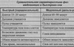
■ In the EEG of a person, waves of electrical activity of different frequencies are observed - from 0.5 to 30 oscillations per second.
Basic rhythms of electrical activity cerebral cortex: alpha rhythm, beta rhythm, delta rhythm and theta rhythm.
Alpha rhythm- vibrations with a frequency of 8-13 hertz; this rhythm prevails over others during sleep.
Beta rhythm has a vibration frequency of more than 13 hertz; it is characteristic of active wakefulness.
Theta rhythm- vibrations with a frequency of 4-8 hertz.
Delta rhythm has a frequency of 0.5-3.5 hertz.
■ Theta and delta rhythms are observed during very deep sleep or anesthesia .
Cranial nerves
Cranial nerves a person has 12 pairs; they depart from different parts of the brain and are divided by function into sensitive, motor and mixed.
❖ Sensory nerves-1, II, VIII pairs:
■ I pair - olfactory nerves that extend from the forebrain and innervate the olfactory region of the nasal cavity;
■ And a couple - visual nerves that branch off from the diencephalon and innervate the retina of the eye;
■ VIII pair - auditory (or vestibule snail f) nerves; depart from the bridge, innervate the membranous labyrinth and the organ of the inner ear.
❖ Motor nerves- III, IV, VI, X, XII pairs:
■ III pair - oculomotor nerves extending from the midbrain;
■ IV pair - blocky nerves also depart from the midbrain;
■ VI - diverting nerves departing from the bridge (III, IV and VI pairs of nerves innervate the muscles of the eyeball and eyelids);
■ XI - additional nerves that extend from the medulla oblongata;
■ XII - sublingual nerves also depart from the medulla oblongata (XI and XII pairs of nerves innervate the muscles of the pharynx, tongue, middle ear, parotid salivary gland).
❖ Mixed nerves-V, VII, IX, X pairs:
■ V pair - trigeminal nerves that extend from the bridge, innervate the scalp, membranes of the eye, chewing muscles, etc.;
■ VII pair - facial nerves, also depart from the bridge, innervate the facial muscles, the lacrimal gland, etc.;
■ IX pair - glossopharyngeal nerves that extend from the diencephalon, innervate the muscles of the pharynx, middle ear, parotid salivary gland;
■ X pair - wandering nerves, also depart from the diencephalon, innervate the muscles of the soft palate and larynx, the organs of the chest (trachea, bronchi, heart, slowing down its work) and abdominal cavities (stomach, liver, pancreas).
Features of the autonomic nervous system
Unlike the somatic nervous system, whose nerve fibers are thick, covered with a myelin sheath and are characterized by a high speed of propagation of nerve impulses, autonomic nerve fibers are usually thin, do not have a myelin sheath and are characterized by a low speed of propagation of nerve impulses (see table). 
❖ Functions of the autonomic nervous system:
■ maintaining the constancy of the internal environment of the body through neuroregulation of tissue metabolism ("launch", correction or suspension of certain metabolic processes) and the work of internal organs, heart and blood vessels;
■ adaptation of the activity of these organs to the changed conditions of the external environment and the needs of the organism.
The autonomic nervous system consists of sympathetic and parasympathetic parts , which have the opposite effect on the physiological functions of organs.
Sympathetic part the autonomic nervous system creates conditions for intensive activity of the organism, especially in extreme conditions, when it is necessary to manifest all the capabilities of the organism.
Parasympathetic part(system "release") of the autonomic nervous system reduces the level of activity, which contributes to the restoration of resources spent by the body.
■ Both parts (divisions) of the autonomic nervous system are subordinate to the higher nerve centers located in hypothalamus , and complement each other.
■ The hypothalamus coordinates the work of the autonomic nervous system with the activity of the endocrine and somatic systems.
■ Examples of the influence of the sympathetic and parasympathetic parts of the ANS on organs are given in the table on p. 520.
Effective performance of the functions of both parts of the autonomic nervous system is ensured double innervation internal organs and heart.
Dual innervation internal organs and the heart means that nerve fibers from both the sympathetic and the parasympathetic parts of the autonomic nervous system are connected to each of these organs. 
The neurons of the autonomic nervous system synthesize various mediators (acetylcholine, norepinephrine, serotonin, etc.) involved in the transmission of nerve impulses.
The main feature autonomic nervous system - bineuronality of the efferent pathway
... This means that in the autonomic nervous system efferent
, or centrifugal
(i.e. coming from the head and dorsal brain to organs
), nerve impulses pass sequentially through the bodies of two neurons. The bi-neuronal nature of the efferent pathway makes it possible to isolate in the sympathetic and parasympathetic parts of the autonomic nervous system central and peripheral parts
.
central part (nerve centers ) the autonomic nervous system located in the central nervous system (in the lateral horns of the gray matter of the spinal cord, as well as in the medulla oblongata and midbrain) and contains the first motor neurons of the reflex arc ... The autonomic nerve fibers that go from these centers to the working organs are switched in the autonomic ganglia of the peripheral part of the autonomic nervous system.
Peripheral part the autonomic nervous system is located outside the central nervous system and consists of ganglion (nerve nodes) formed by bodies second motor neurons of the reflex arc as well as nerves and nerve plexuses.
■ Have sympathetic division, these ganglia form a pair sympathetic chains (trunks), located near the spine on both sides, in the parasympathetic section, they lie near or inside the innervated organs.
■ Postganglionic parasympathetic fibers are suitable for the eye muscles, larynx, trachea, lungs, heart, lacrimal and salivary glands, muscles and glands of the digestive tract, excretory and genitals. 
Causes of impaired activity of the nervous system
❖ Overwork of the nervous system weakens its regulatory function and can provoke the emergence of a number of mental, cardiovascular, gastrointestinal, skin and other diseases.
❖ Hereditary diseases can lead to a change in the activity of some enzymes. As a result, toxic substances accumulate in the body, the effect of which leads to impaired development of the brain and mental retardation.
Negative environmental factors:
■bacterial infections lead to the accumulation of toxins in the blood, poisoning the nervous tissue (meningitis, tetanus);
■ viral infections can affect the spinal cord (poliomyelitis) or the brain (encephalitis, rabies);
■ alcohol and products of its exchange excite various nerve cells (inhibitory or excitatory neurons), disorganizing the work of the nervous system; systematic alcohol consumption causes chronic depression of the nervous system, changes in skin sensitivity, muscle pain, weakening and even disappearance of many reflexes; irreversible changes occur in the central nervous system, forming personality changes and leading to the development of severe mental illness and dementia;
■ influence nicotine and drugs much the same as alcohol;
■heavy metal salts bind to enzymes, disrupting their work, which leads to disturbances in the activity of the nervous system;
■ at poisonous animal bites biologically active substances (poisons) enter the bloodstream, disrupting the functioning of neuronal membranes;
■ at head trauma, bleeding, and severe pain loss of consciousness is possible, which is preceded by: darkening in the eyes, tinnitus, pallor, lowering temperature, profuse sweat, weak pulse, shallow breathing.
Cerebral circulation disorder. Narrowing of the cerebral lumen leads to disruption of the normal functioning of the brain and, as a consequence, to diseases of various organs. Injury and high blood pressure can cause rupture of blood vessels in the brain, which usually leads to paralysis, high nerves, or death.
Compression of the nerve stems of the brain causes severe pain. Infringement of the spinal cord roots by spasmodic muscles of the back or as a result of inflammation causes paroxysmal pain (characteristic of radiculitis ), violation of sensitivity ( numbness ) and etc.
❖ When metabolic disorders in the brain mental illness occurs:
■neurosis - emotional, motor and behavioral disorders, accompanied by deviations from the autonomic nervous system and the work of internal organs (example: fear of the dark in children);
■ affective insanity - A more serious illness in which periods of extreme excitement alternate with apathy (paranoia, megalomania or persecution);
■ schizophrenia - splitting of consciousness;
■ hallucinations (can also occur in case of poisoning, high temperature, acute alcoholic psychosis).
The human body is a multistage structure, each organ and system of which is closely interconnected with each other and with the environment. And so that this connection is not interrupted for a split second, a nervous system is provided - a complex network that permeates the entire human body and is responsible for self-regulation and the ability to adequately respond to external and internal stimuli. Thanks to the well-coordinated work of the nervous system, a person can adapt to the factors of the external world: any, even insignificant, change in the environment forces nerve cells to transmit hundreds of impulses at an incredibly high speed so that the body can instantly adapt to new conditions for itself. Internal self-regulation works in a similar way, in which the activity of cells is coordinated in accordance with current needs.
The functions of the nervous system affect the most important life processes, without which the normal existence of the organism is unthinkable. These include:
- regulation of the work of internal organs in accordance with external and internal impulses;
- coordination of all units of the body, from the smallest cells to organ systems;
- harmonious human interaction with the environment;
- the basis of higher psychophysiological processes inherent in humans.
How does this complex mechanism work? What cells, tissues and organs is the human nervous system represented and what is each of its departments responsible for? A short excursion into the basics of the anatomy and physiology of the human body will help find answers to these questions.
Organization of the human nervous system
Nerve cells cover the entire body as a whole, forming an extensive network of fibers and endings. This system, on the one hand, unites every cell of the body, forcing it to work in one direction, and on the other hand, it integrates a particular person into the environment, balancing his needs with external factors. The nervous system ensures the normal processes of digestion, respiration, blood circulation, the formation of immunity, metabolism, etc. - in a word, everything without which normal life is inconceivable.
The effectiveness of the nervous system depends on the correct formation of the reflex - the response of the body to irritation. Any impact, be it external changes or internal imbalance, triggers a chain of impulses that instantly affect the body, and he, in turn, forms a response. Thus, the human nervous system forms the unity of tissues, organs and systems of the human body with each other and with the surrounding world.
The entire nervous system consists of millions of nerve cells - neurons, or neurocytes, each of which has a body and several processes.
The classification of neuron processes depends on what function it performs:
- the axon sends a nerve impulse from the body of the neuron to another nerve cell, or the ultimate goal of the chain is a tissue or organ that must perform a certain action;
- the dendrite receives the sent impulse and leads it to the body of the neuron.
Due to the fact that each nerve cell is polarized, the chain of nerve impulses never changes direction, getting back on track. Thus, each nerve impulse is promoted, initiating the work of muscles, internal organs and systems.
Varieties of nerve cells
Before considering the nervous system as a whole, it is necessary to understand what functional units it consists of. The National Assembly includes:
- Sensitive neurons. They are located in nerve nodes that receive information directly from receptors.
- Intercalary neurons are an intermediate link through which the received impulse is transmitted from sensory neurons further along the chain.
- Motor neurons. They act as initiators of a response to a stimulus, transmitting a signal from the brain to the muscles or glands, which normally should perform the function assigned to them.

It is according to this scheme that any response of the human body to an external or internal signal-stimulus, which acts as an impetus for a specific action, is built. As a rule, the passage of a nerve impulse takes a few fractions of a second, but if this time is delayed or the chain is interrupted, this indicates the presence of a pathology of the nervous system and requires serious diagnosis.
The structure and types of the nervous system: structural classification
To simplify the structure of the nervous system, in medicine, there are several classifications depending on the structure and functions performed. So, anatomically, the human nervous system can be divided into 2 broad groups:
- central (CNS), formed by the brain and spinal cord;
- peripheral (PNS), represented by nerve nodes, endings and nerves directly.
The basis of this classification is extremely simple: the central nervous system is a kind of connecting link in which the incoming impulse is analyzed and further regulation of the activity of organs and systems is carried out. And the PNS serves to transport the incoming signal from the receptors to the central nervous system and the subsequent activator, but from the central nervous system to the cells and tissues that will perform a specific action.
central nervous system
The central nervous system is a key component of the nervous system, because it is here that the main reflexes are formed. It consists of a spinal cord and a brain, each of which is reliably protected from external influences by bone structures. Such thoughtful protection is necessary, since each part of the central nervous system performs vital functions, without which it is impossible to maintain health.
Spinal cord
This structure is enclosed within the spinal column. She is responsible for the simplest reflexes and involuntary reactions of the body to a stimulus.

In addition, neurons in the spinal cord coordinate the activity of muscle tissue, which regulates defense mechanisms. For example, having felt an extremely hot temperature, a person involuntarily pulls his palm, thereby protecting himself from thermal burns. This is a typical spinal cord-controlled response.
Brain
The human brain consists of several sections, each of which performs a number of physiological and psychological functions:
- The medulla oblongata is responsible for the vital functions of the body - digestion, breathing, the movement of blood through the vessels, etc. In addition, the nucleus of the vagus nerve is located here, which regulates the autonomic balance and psychoemotional response. If the nucleus of the vagus nerve sends active impulses, the vitality of a person decreases, he becomes apathetic, melancholic and depressive. If the activity of impulses emanating from the core decreases, the psychological perception of the world changes to a more active and positive one.
- The cerebellum regulates the precision and coordination of movements.
- The midbrain is the main coordinator of muscle reflexes and tone. In addition, neurons regulated by this part of the central nervous system contribute to the adaptation of the sense organs to external stimuli (for example, accommodation of the pupil at dusk).
- The diencephalon is formed by the thalamus and hypothalamus. The thalamus is the most important organ-analyzer of incoming information. In the hypothalamus, the emotional background and metabolic processes are regulated, there are centers responsible for the feeling of hunger, thirst, fatigue, thermoregulation, and sexual activity. Thanks to this, not only physiological processes are coordinated, but also many human habits, for example, the tendency to overeat, the perception of cold, etc.
- The cortex of the cerebral hemispheres. The cerebral cortex is a key link in mental functions, including consciousness, speech, perception of information and its subsequent comprehension. The frontal lobe regulates motor activity, the parietal is responsible for bodily sensations, the temporal lobe controls hearing, speech and other higher functions, and the occipital contains the centers of visual perception.

Peripheral nervous system
The PNS provides interconnection between organs, tissues, cells and the central nervous system. Structurally, it is represented by the following morphological and functional units:
- Nerve fibers, which, depending on the functions performed, are motor, sensitive and mixed. The motor nerves transmit information from the central nervous system to the muscle fibers, the sensitive ones, on the contrary, help to perceive the information received with the help of the senses and transmit it to the central nervous system, and the mixed ones, to one degree or another, participate in both processes.
- Nerve endings, which are also motor and sensitive. Their function is no different from fiber structures with a single caveat - nerve endings begin or, conversely, end a chain of impulses from organs to the central nervous system and vice versa.
- Nerve nodes, or ganglia, are clusters of neurons outside the central nervous system. The spinal ganglia are responsible for the transmission of information received from the external environment, and the vegetative - data on the state and activity of internal organs and resources of the body.
In addition, all peripheral nerves are classified according to their anatomical features. Based on this characteristic, 12 pairs of cranial nerves are distinguished, which coordinate the activity of the head and neck, and 31 pairs of spinal nerves responsible for the trunk, upper and lower extremities, as well as internal organs located in the abdominal and chest cavities.
The cranial nerves originate from the brain. The basis of their activity is the perception of sensory impulses, as well as partial participation in respiratory, digestive and cardiac activity. The function of each pair of cranial nerves is presented in more detail in the table.
| P / p No. | Name | Function |
| I | Olfactory | Responsible for the perception of various odors, transmitting nerve impulses from the olfactory organ to the corresponding center of the brain. |
| II | Visual | Regulates the perception of visual data by delivering impulses from the retina. |
| III | Oculomotor | Coordinates the movement of the eyeballs. |
| IV | Block | Along with the oculomotor pair of nerves, it takes part in the coordinated mobility of the eyes. |
| V | Trigeminal | Responsible for sensory perception of the facial area, and also participates in the act of chewing food in the oral cavity. |
| VI | Diverting | Another nerve that regulates the movement of the eyeballs. |
| Vii | Facial | The nerve that coordinates facial contractions of the facial muscles. In addition, this pair is also responsible for gustatory perception, transmitting signals from the papillae of the tongue to the brain center. |
| VIII | Vestibular cochlear | This pair is responsible for the perception of sounds and the ability to maintain balance. |
| IX | Glossopharyngeal | Regulates the normal activity of the pharyngeal muscles and partially transmits taste sensations to the brain center. |
| X | Wandering | One of the most important cranial nerves, the functionality of which depends on the activity of the internal organs located in the neck, chest and abdominal walls. These include the pharynx, larynx, lungs, heart muscle and organs of the digestive tract. |
| XI | Dorsal | Responsible for the contraction of muscle fibers of the cervical and shoulder regions. |
| XII | Sublingual | Coordinates the activity of the language and partially forms the speech skill. |
The activity of the spinal nerves is classified much more simply - each specific pair or complex of pairs is responsible for the section of the body of the same name assigned to it:
- cervical - 8 pairs,
- breast - 12 pairs,
- lumbar and sacral - 5 pairs, respectively,
- coccygeal - 1 pair.
Each member of this group belongs to mixed nerves formed by two roots: sensory and motor. That is why the spinal nerves can perceive an irritating effect, transmitting an impulse along a chain, and activate activity in response to a message from the central nervous system.

Morphofunctional division of the nervous system
There is also a functional classification of parts of the nervous system, which includes:
- Somatic nervous system, which regulates the functions of skeletal muscles. It is controlled by the cerebral cortex, therefore it is completely subordinated to the conscious decisions of a person.
- The autonomic nervous system is responsible for the activity of internal organs. Its centers are located in the brain stem, and therefore it is not consciously regulated in any way.
In addition, the autonomic system is further subdivided into 2 significant functional divisions:
- Sympathetic. It is activated when energy is expended;
- Parasympathetic. Responsible for the recovery period of the body.

Somatic nervous system
Somatics is a division of the nervous system that is responsible for the delivery of motor and sensory impulses from receptors to the organs of the central nervous system and vice versa. Most of the nerve fibers in the somatic system are concentrated in the skin, muscle frame and organs responsible for sensory perception. It is the somatic nervous system that almost 100% coordinates the conscious part of the activity of the human body and the processing of information received from the receptors of the sense organs.
The main elements of somatics are 2 types of neurons:
- sensory, or afferent. Regulate the delivery of information to the cells of the central nervous system;
- motor, or efferent. They work in the opposite direction, transporting nerve impulses from the central nervous system to cells and tissues.
Both those and other neurons stretch from the central nervous system directly to the final target of the impulses, that is, to muscle and receptor cells, and the body in most cases is located directly in the central part of the nervous system, and the processes reach the required localization.
In addition to conscious activity, the somatic also includes a part of reflexes that are unconsciously controlled. With the help of such reactions, the muscular system comes into an active state without waiting for an impulse from the brain, which allows it to act instinctively. Such a process is possible if the pathways of the nerve fibers pass directly through the spinal cord. An example of such actions is twitching the hand when a high temperature is felt or a knee reflex when hitting a tendon with a hammer.
Autonomic nervous system
Vegetation, or the autonomic nervous system, is a department that coordinates the activity of mainly internal organs. Since the main life processes - respiration, metabolism, heart contractions, blood flow, etc. - are not subject to consciousness, autonomic nerve fibers react mainly to changes in the internal environment of the body, remaining indifferent to conscious impulses. Thanks to this, the body maintains optimal conditions for providing the energy resources necessary in a particular situation.

The features of autonomic nervous activity imply that the main fibers are concentrated not only in the organs of the central nervous system, but also in other tissues of the human body. Numerous nodes are scattered throughout the body, forming the autonomic nervous system outside the central nervous system, between the brain centers and organs. Such a network can regulate the simplest functions, but more complex mechanisms still remain under the direct control of the central nervous system.
The key role of vegetation is to maintain a relatively constant homeostasis by self-tuning the activity of internal organs, depending on the needs of the body. So, vegetative fibers optimize the secretion of hormones, the rate and intensity of blood supply to tissues, the intensity and frequency of respiration and heart rate, and other key mechanisms that must respond to changes in the external environment (for example, during intense physical activity, an increase in temperature or humidity, atmospheric pressure, etc. etc.). Thanks to these processes, compensatory and adaptive reactions are provided that maintain the body in optimal shape under any circumstances. Since the unconscious activity of internal organs can be regulated in two directions (activation and suppression), vegetation can also be conditionally divided into 2 sections - parasympathetic and sympathetic.
Sympathetic nervous system
The sympathetic part of the vegetation is directly connected with the spinal substance located from the first thoracic to the third lumbar vertebra. It is here that the stimulation of the activity of internal organs is carried out, which is necessary during increased energy consumption - during physical exertion, during stress, intense work or emotional shock. Such mechanisms make it possible to support the body, providing it with the resources necessary to overcome adverse conditions.
Under the influence of sympathy, respiration and vascular pulsation become more frequent, due to which the tissues are better supplied with oxygen, energy is released from the cells faster. Thanks to this, a person can work more actively, coping with increased loads in conditions of disadvantage. However, these resources cannot be infinite: sooner or later, the amount of energy reserves decreases, and the body can no longer function "at increased speed" without respite. Then the parasympathetic department of vegetation is included in the work.
Parasympathetic nervous system
The parasympathetic nervous system is localized in the midbrain and sacral spine. She, unlike sympathizers, is responsible for maintaining and accumulating energy depots, reducing physical activity and good rest.
So, for example, parasympathetic slows down the heart rate during sleep or physical rest, when a person recovers spent strength, coping with fatigue. Additionally, at this time, peristaltic processes are activated, which have a positive effect on metabolism and, as a result, on the restoration of nutrient reserves. Thanks to such self-regulation, defense mechanisms are activated, which are especially important at a critical level of overwork or exhaustion - the human body simply refuses to continue working, requiring time for rest and recovery.
Features and differences of the sympathetic and parasympathetic nervous system
At first glance, it may seem that the sympathetic and parasympathetic divisions are antagonists, but in reality this is not so. Both of these departments act in a coordinated manner and together, simply in different directions: if the sympathetic activates the work, then the parasympathetic allows you to recover and rest. Thanks to this, the work of the internal organs always, to a greater or lesser extent, corresponds to a specific situation, and the body can adapt to any conditions. In fact, both of these systems form the basis of homeostasis, balancing the activity levels of the human body.
Most of the internal organs have both sympathetic and parasympathetic fibers, which have different effects on them. Moreover, the state of the organ at the current moment depends on which of the departments of the National Assembly prevails in the current circumstances. An illustrative example of the activity of these systems can be seen in the table below.
| Organ | Parasympathetic effects | Sympathetic impact |
| Blood supply to the brain | Constriction of blood vessels, decrease in the volume of incoming blood | Expansion of blood vessels, activation of blood supply |
| Peripheral arteries and arterioles | Narrowing of the lumen, increasing blood pressure and weakening blood flow | Expansion of the diameter of arterial vessels and decrease in pressure |
| Heart rate | Decrease in heart rate | Increased heart rate |
| Digestive system | Strengthening gastrointestinal motility for faster absorption of nutrients | Slowdown of peristalsis and, as a result, metabolism |
| Salivary glands | Increased secretion | Feeling of dry mouth |
| Adrenal glands | Suppression of endocrine function | Activation of hormone synthesis |
| Bronchi | Narrowing of the bronchial lumen, heavier unproductive breathing | Expansion of the bronchi, an increase in the volume of inhaled air and the productivity of each respiratory movement |
| Visual analyzer | Constriction of the pupils | Dilated pupils |
| Bladder | Reduction | Relaxation |
| Sweat glands | Decreased sweating | Increased activity of sweat glands |
Post Scriptum
Neurological problems associated with diseases of the human nervous system are among the most difficult in medical practice. Any damage to the nerve tissue leads to a partial or complete loss of control over the body, causing tremendous damage to the quality of life and reducing a person's functionality. Only a complex and coordinated action of each neuron in all parts of the central and peripheral NS is able to maintain the body in an optimal state, ensure the correct operation of each organ, adequately fit into the surrounding realities and respond to external stimuli. Therefore, it is necessary to carefully monitor the health of your own nervous system, and at the slightest suspicion of a deviation, urgently take appropriate measures - this is one of those cases in which it is better to engage in prevention than to waste time while it is still possible to fix it without consequences!