"Hereditary diseases" - Intercurrent diseases worsen the condition of patients. Symptoms: from severe mental retardation to the complete absence of any symptoms. The peak of the disease occurs at 2 years. The diagnosis is usually established after 3 years, when the subluxation of the lens is revealed. Decreased intelligence is detected only in 50% of sick children.
Coma - Breathing slows down. Mixed with raw or processed opium for later use. Intoxication - (from the Latin prefix in - "in" and the Greek toxikon - poison). Pathogenesis of uremic coma. Disorders of mental activity (depression, motor restlessness, emotional lability) are typical.
"Kidney Disease" - The main symptoms of kidney disease. Acceptance of spicy dishes and seasonings is limited. In general, women predominate among patients with pyelonephritis. Inflammation of the kidneys Pyelonephritis. Kidney disease. A dairy-vegetable diet is recommended, meat and boiled fish are allowed. The process of formation and excretion of urine is called diuresis.
"Genetic Disorders" - DNA analysis revealed traces of hemophilia. Russia was no exception. Types of gene mutations. The likelihood of heredity. Hemophilia is a hereditary disease characterized by a violation of the blood coagulation mechanism. Many descendants of Queen Victoria suffered from the disease. Human genetic diseases are inherited.
"Apnea" - Decreased tone of the pharyngeal muscles. In 2001, there were about 10 sleep laboratories in Russia. Sleep disorder. Patient G., 52 years old, severe OSAS. Obesity 2-4 tbsp. The characteristic appearance of a patient with obstructive sleep apnea syndrome. CPAP therapy (Continuous Positive Airway Pressure). Laser plastic surgery of the sky.
"Gastrointestinal diseases" - 5. Hepatic flukes. 2. You need to eat raw vegetables and fruits. 3. 5. At lunch, it is important to eat the salad first, and then the soup. Food hygiene rules. Helminthic diseases. 6. Food poisoning. Ascaris.
There are 27 presentations in total

2 Etiology of coma The causes of coma are very diverse. They can be due to many exogenous and endogenous influences on the body. The variety of etiological causes of coma, however, leads to universal pathophysiological and clinical aspects of this problem. But coma in various diseases have their own pathogenetic and clinical features. In some cases, irreversible changes against the background of a coma develop rapidly, in others there is a uniformity of pathophysiological mechanisms in the development of coma with a gradual transition of functional changes into irreversible organic ones. The development of coma is most often caused by: poisoning, craniocerebral trauma, infectious diseases (sepsis, meningoencephalitis), brain tumors, complications of endocrine diseases (diabetes mellitus, etc.), liver and kidney diseases, impaired temperature homeostasis, posthypoxic encephalopathies.

3 Pathophysiological aspects of coma development In all diseases, the basis of the pathogenesis of coma is damage to the central nervous system, the degree of damage to which largely depends on the prognosis of the restoration of body functions. The leading factors in the development of a coma are disorders of cerebral circulation, disorders of cerebrospinal fluid circulation, hypoxia, acidosis, as a result of which edema of the cerebral cortex occurs, against the background of which there is an energy starvation of the brain associated with insufficient supply of oxygen, nutrient substrates or a violation of their utilization in case of toxic damage to the nerves. cells. Loss of consciousness is always associated with a disruption in the activity of the cerebral cortex of the primary or secondary plan. With the latter, the functions of the brain stem are first disturbed, which entails loss of consciousness as a result of the secondary shutdown of the activity of the cortex. Impairment of consciousness in lesions of the brain stem is due to the disintegration of the reticular formation, and in metabolic disorders - by a diffuse effect on the reticular formation or both hemispheres. The peculiarities of the propagation of the phases of inhibition and excitation in the cerebral cortex and subcortex lead to a disturbance of consciousness of a certain depth.

4 Examination of a patient in a coma - 1 Admission of a patient in a coma, identification of the cause and type of coma are significant difficulties, therefore, as soon as the patient's vital functions are preserved and the severity of the patient's condition according to the Glasgow scale is determined, it is necessary to start taking anamnesis and examining the patient. Anamnestic data need to be obtained from relatives, friends, police officers, persons who took the patient to the hospital, to find out the circumstances of the coma development, the diseases that preceded it: did the patient have diabetes, epilepsy, kidney disease. An examination of the patient's belongings is necessary, where you can find medical documents (certificate of a patient with diabetes mellitus), as well as medications. Focus on finding out the onset of the disease. Coma onset (sudden, gradual), recent complaints (headache, depression, focal weakness, dizziness). Recent trauma, previous illnesses (diabetes, uremia). Anamnestic data on previous psychiatric disorders. Access to medicines. A sudden onset coma, caused by an unknown cause in a previously healthy person, is most often caused by self-poisoning with drugs, skull trauma, subarachnoid or brain stem hemorrhage. With metabolic comas, impairment of consciousness usually develops gradually. On examination, it is necessary to undress the patient completely, look for signs of trauma, acute or chronic general illness, injection or self-poisoning with drugs.

5 Examination of a patient in a coma - 2 Pay attention to the color of the skin, mucous membranes, odor from the mouth, the size of the pupils, their asymmetry and slowing down of reaction to light, examine the fundus. The character of spontaneous movements, muscle tone, the presence of seizures of pathological reflexes are assessed. Determine the frequency and type of breathing. Listen and examine the boundaries of the heart, blood pressure, the size of the liver, spleen. Laboratory tests are prescribed in accordance with the history and objective research data. CBC, sugar level, acid-base state and gas composition of capillary or arterial blood are determined. Prothrombin time, bilirubin, transaminase activity. The content of potassium, sodium, magnesium, blood urea nitrogen, creatinine. Toxicological screening of blood and urine is carried out. In addition to the clinical analysis of urine, the presence of sugar, ketone bodies is determined there. X-rays of the skull and cervical spine are necessary for possible injury. Electroencephalography establishes the epileptogenic nature of the coma. Computed tomography and nuclear magnetic resonance are indicated for hemorrhages and masses of the brain. Lumbar puncture is performed with a certain clinical picture. Treatment The general principles of therapy in coma are the maintenance of vital functions and specific treatment depending on the cause of the coma.

6 Coma in diabetes mellitus in children Insulin-dependent diabetes mellitus (IDDM) is more common in children. With IDDM, the development of a coma can be with insulin deficiency, dietary disorders, concomitant infectious diseases, mental or physical trauma. In this regard, the symptoms of diabetes often remain in the shadows, the child is treated for an infectious disease against the background of ongoing decompensation of diabetes. The child can be hospitalized with a diagnosis of acute abdomen, acetonemic vomiting, meningitis. Therefore, a study of blood sugar is necessarily shown in the serious condition of a child, especially when entering the intensive care unit. There are the following types of diabetic coma: Ketoacidotic, Hyperosmolar, Hyperlactatemic (lactic acid), Hypoglycemic.

7 Ketoacidotic coma - 1 Ketoacidotic coma occurs most often in children and is characterized by its gradual development. The diagnosis of ketoacidotic coma is made on the basis of anamnesis and a combination of clinical and biochemical data. Only insufficient knowledge of the clinic of diabetes mellitus can lead to its late diagnosis. The clinical sign of incipient ketoacidosis is increasing thirst and polyuria, weight loss, decreased appetite, nausea, dry skin and mucous membranes, and acetone odor from the mouth. At the first stage, these symptoms are joined by a sharp weakness, constant drowsiness. Skin and tendon reflexes are reduced. There is vomiting and abdominal pain syndrome. With the deepening of the coma - the second stage - the impairment of consciousness increases. The child can be woken up and he will answer monosyllabic questions. Breathing is noisy, deep - Kussmaul. Vomiting, abdominal pain intensifies. The third stage of coma is impaired consciousness against the background of an extremely serious condition. Tendon and skin reflexes are absent. Symptoms of dehydration, against the background of tachycardia, threadlike pulse, oligoanuria. Coma manifestation can be manifested by the predominance of one of two syndromes: abdominal or cardiovascular. The abdominal syndrome usually develops rapidly, which is often an indication for emergency hospitalization of a child with a diagnosis of acute abdomen, appendicitis. Thirst, polyuria, weight loss, nausea, vomiting are characteristic. Soreness and tension in the muscles of the abdomen.

8 Ketoacidotic coma - 2 Cardiovascular syndrome is determined by symptoms of heart failure: threadlike pulse, low blood pressure, cold, pale cyanotic skin, oliguria. The child may have heart pain, dizziness, fainting. A laboratory study reveals: hyperglycemia, a decrease in reserve alkalinity, acidosis (a decrease in blood pH), ketonemia, glucosuria, ketonuria, hypocapnia, hypoxemia, an increase in lactate and pyruvate. Hyponatremia - in children with abdominal syndrome and sodium - at the upper limit of the norm with a cardiovascular variant of the coma. The osmolarity of blood is up to extremely high (350 mosm / l) of its figures, which determines the tactics of choosing starting infusion solutions to increase or decrease the osmolarity of blood to the upper limits of the norm by the twelfth hour of treatment. This accelerates the removal of patients from a coma and minimizes the risk of developing cerebral edema, as the most formidable complication of inadequate therapy.

9 Hyperosmolar coma Hyperlactatemic (lactic acid) coma Hyperosmolar coma is a sharp increase in blood osmolarity against a background of high glycemia (above mmol / L) and hypernatremia, above 150 mmol / L, as well as chloremia, increased urea levels. In this case, there is no ketoacidosis. Other laboratory parameters are also changed: a high level of hemoglobin and hematocrit value of blood, leukocytosis. High sugar content in urine. Blood pH and bicarbonate levels are normal. No hyperketonemia. The reaction to acetone in urine is negative. The clinical feature of hyperosmolar coma is rapidly developing severe symptoms of dehydration - dry skin, mucous membranes, decreased turgor of the eyeballs. Unlike ketoacidosis, there is no smell of acetone in the exhaled air. Disorder of consciousness - from drowsiness to deep coma. There may be pathological reflexes and hyperthermia. Superficial rapid breathing, tachycardia, decreased blood pressure. Oligoanuria. The development of hyperosmolar coma often provokes excessive intake of carbohydrates, intercurrent diseases. The classic variant of the course of hyperosmolar coma in children is rare. Hyperlactatemic (lactic acid) coma, as a rule, develops with an increase in anaerobic glycolysis against the background of hypoxia and the associated accumulation of lactic acid. Biochemical changes lead to characteristic clinical symptoms: agitation, aggressiveness, increased motor restlessness of the child, shortness of breath. Pains are observed in the region of the heart and behind the sternum, muscles of the upper and lower extremities. A laboratory study reveals moderate hyperglycemia, a sharp shift in the acid-base state to the acidic side, hyperpyruvatemia. The diagnostic feature of this type of coma is the absence of ketosis: ketone bodies in the urine and exhaled air, which sometimes complicates the diagnosis of IDDM in young children.

10 Principles of therapy for diabetic coma - 1 1. Children in a coma need the following laboratory tests: determination of the level of glycemia, the acid-base state of the blood, the level of hemoglobin and hematocrit, serum electrolytes (potassium, sodium, calcium, phosphorus, chlorine), the level of blood urea and transaminases. Coagulograms, clinical analyzes of blood, urine, sugar and acetone in urine, lactate, pyruvate and ECG. 2. The study of glycemia, CBS, electrolytes should be repeated every 3-4 hours. 3. Emergency therapy in the early manifestations of coma and in ketoacidosis should begin with gastric lavage with 2% sodium bicarbonate solution, in case of abdominal syndrome - with saline, as well as a cleansing enema. 4. Infusion of sodium bicarbonate solution should be carried out only with a pronounced shift in blood pH to the acidic side (pH

11 Principles of therapy for diabetic coma - 2 Fluid infusion is recommended with solution 1, used as a starting point - in children with cardiovascular syndrome, and solution 2, used more in patients with abdominal syndrome (V.V. Smirnov, 1998) ... It is recommended to alternate solutions 1 and 2 in a 1: 1 ratio. Hyperosmolar coma involves the initiation of infusion therapy with a transfusion of 0.45% (hypotonic sodium chloride solution). The subsequent infusion of fluid is continued with solutions 1 and 2. Solution 2 Ringer's solution 200.0 Glucose solution 40% KCl4 solution 5% Insulin 3-6 units. Heparin UNITS Panangin solution 10.0 Solution 1 Glucose solution 5-10% KCl4-5% solution Insulin 3-6 units Heparin ED KKB mg

12 Principles of therapy for diabetic coma - 3 7. The level of glycemia in a comatose patient must be maintained within mmol, without reducing it to mmol / l and below to avoid hypoglycemia. The decrease in high glucose concentration should be gradual, no more than 2 times in 8-12 hours. Since a rapid decrease in the level of glycemia can also lead to hypoglycemia with a worsening of the patient's condition. 8. Maintaining the level of glycemia within 15.5 mmol / l is achieved by changing the ratio of glucose / insulin in the injected solution: Glycemia, mmol / l Glucose Insulin, units Glucose / Insulin%%%%%%%%

13 Principles of therapy for diabetic coma - 4 9. Administration of potassium, panangin, Ringer's solution is indicated 3 hours after the start of infusion therapy. 10. A child with a coma is shown the obligatory prescription of antibiotics of a wide range of action, possibly two, to suppress all clinically significant pathogens. 11. Heparin therapy is carried out at the rate of U / kg of body weight per day. 12. For the prevention of cerebral edema, children are prescribed a 25% solution of magnesium sulfate - 0.2 ml / kg of body weight per day. 13. In case of heart failure, cardiac glycosides, dopamine, dobutrex, glucocorticoids are prescribed. 14. Diet therapy for a child in a coma. Nutrition for a patient in a coma is prescribed only when the condition improves, at the beginning in the form of drinking mineral water, tea, compote. During ketosis, butter is completely excluded. Improvement of clinical symptoms implies an increase in the diet. Calculation of the dose of insulin in accordance with the amount of food eaten, measured in bread units.

14 Hypoglycemic coma Hypoglycemic coma is caused by a decrease in the level of glucose in the blood, accompanied by a rapid loss of consciousness due to an overdose of insulin, missed meals, and heavy physical exertion. The initial symptoms of hypoglycemia are hunger, weakness, cold sweat, headache, restlessness, and sometimes drowsiness. In newborns and infants, hypoglycemia is manifested by anxiety, unmotivated crying, and aggressive behavior. Against the background of a lack of consciousness in hypoglycemic coma, profuse sweat, moist mucous membranes, convulsions, trismus of the masticatory muscles, Babinsky's symptoms are noted. Hypoglycemic coma requires its differentiation from other types of diabetic coma, epilepsy. With the initial symptoms of hypoglycemia, it is necessary to give the child a sweet drink (tea, juice, sugar syrup), easily digestible carbohydrates food (jam, honey, candy, porridge, white bread). With the development of hypoglycemic coma, 20, 40, 60 ml of a 40% glucose solution are injected intravenously until consciousness returns. Then the fluid is infused with solution 1, where no insulin is added, and with solution 2 under the control of blood sugar levels. Along with the introduction of glucose, in some cases, glucagon is introduced, which promotes the conversion of liver glycogen into glucose. Glucagon is administered at a dose of 0.5-1.0 ml intramuscularly or subcutaneously.

15 Coma in traumatic brain injury - 1 Severe traumatic brain injury is accompanied by the development of a coma in 10-20% of cases. The most common causes of severe damage to the skull and brain are transport injuries, as well as falls from a height, blows to the head with hard objects. Often, a gross disturbance of consciousness occurs after a "light" interval, during which there may be stunning, drowsiness or psychomotor agitation. "Bright" gap - as evidence of progressive compression of the brain by intracranial hematoma or is associated with increasing cerebral edema. With severe bruises of the trunk-basal sections, the coma can last up to several weeks. In a coma in patients, cerebral symptoms prevail. Vomiting is a mandatory symptom of severe trauma. It occurs immediately or after 1-2 hours after injury. Myiasis or mydriasis is determined, which in the absence of a photoreaction serves as an unfavorable prognostic sign. Patients have ptosis, strabismus, floating movements and uneven standing of the eyeballs. Corneal reflexes are absent, spontaneous horizontal nystagmus. Bilateral increase in muscle tone in the limbs. Paresis and paralysis can have the character of tetra- and monohemiparesis. Pathological reflexes of Babinsky, Oppenheim, oral automatism, Kernig, Brudzinsky, and stiff neck appear.

16 Coma in traumatic brain injury - 2 Pathological forms of respiration such as Cheyne-Stokes, Biota, terminal with separate breaths and subsequent apnea. Possibly tachypnea. With the retraction of the tongue, lower jaw, aspiration of blood, stomach contents - breathing is frequent, noisy, snoring, with the participation of auxiliary muscles. Blood pressure changes either towards hypotension or hypertension. The rhythm of the heartbeats changes. The most common is tachycardia, but bradycardia is also possible. Hyperthermia - in the first hours, sometimes 1-2 days after the injury. The most important moment that determines the course of the disease in severe traumatic brain injury is the syndrome of compression of the brain, the presence of which requires immediate surgical intervention. The compression syndrome is manifested by a deepening coma, an increase in meningeal symptoms, the appearance of seizures, mono- and hemiparesis. The most common cause of compression syndrome is epi- and subdural hematomas. With intraventricular hematomas, there are harmetonic crises, autonomic disorders. Compression of the brain develops with its dislocation and compression of the brainstem sections. Disorder of vital functions quickly sets in. For a fracture of the base of the skull, hemorrhages around the eyes (glasses) are characteristic. Bleeding and liquorrhea from the nose, external auditory canal, nasopharynx and lesions of the cranial nerves are also noted.

17 Special research methods Lumbar puncture is performed in a patient in a shallow coma. In a deep coma and with suspicion of intracranial hematoma, lumbar puncture is contraindicated. In a coma with traumatic brain injury, there can be either an increase in CSF pressure or a decrease. The composition of the cerebrospinal fluid in patients without subarachnoid hemorrhage is normal in the first days after injury, but later some cytosis and an increase in protein content are noted. With subarachnoid hemorrhage, an admixture of blood is found. ECHO-EG - is a valuable study that helps to establish or with a degree of probability to reject the presence of intracranial hemorrhage. In children in a deep coma, there may be a disappearance or a sharp weakening of the pulsation of echo signals. On the EEG in a coma with traumatic brain injury, a violation of the regular rhythm and interhemispheric asymmetry are noted with bruises or hematomas. Angiography is an important diagnostic method for identifying intracranial and intracerebral hematomas. Radioisotope and ultrasound research methods, computed tomography and nuclear magnetic resonance imaging of the brain are very informative for the diagnosis of traumatic brain injury in children.

18 Intensive therapy for coma associated with traumatic brain injury Treatment of children with traumatic brain injury should begin with the correction of impaired vital functions. This is, first of all, the restoration of breathing and the maintenance of hemodynamics. Provide airway patency, carry out oxygen therapy, if necessary - artificial ventilation of the lungs. In the correction of hemodynamic disorders, first of all, the volume of circulating blood is replenished: blood is transfused, blood substitutes (rheopolyglucin, albumin), against the background of the introduction of cardiotonic drugs - dopamine, dobutrex. Surgical intervention is carried out simultaneously with the correction of vital functions. Dehydration is an essential component of intensive treatment. For this purpose, the administration of lasix at a dose of 4-5 mg / kg of body weight per day and / or intravenous infusion of mannitol at a dose of 1 g / kg of body weight is used. Inside, glycerin is used - 1 g / kg of body weight. In severe cerebral edema, dexamethasone mg / kg of body weight per day is prescribed. Lytic mixtures are introduced containing antihistamines, neuroplegic and ganglion blocking drugs: suprastin, glucose-novocaine mixture (0.25% novocaine solution together with an equal amount of 5% glucose). To stop hyperthermia, a 25-50% solution of analgin is used, physical methods of cooling (cold on the main vessels, fans, cold wraps). To improve cerebral hemodynamics include aminophylline, no-shpa, trental (pentoxifylline), curantil (dipyridamole). Hemostatic drugs are used - vicasol, calcium chloride, dicinone, protease inhibitors - contrikal, gordox. Broad-spectrum antibiotics are prescribed. Convulsive syndrome is stopped by the introduction of GHB, seduxen. Restoration of brain functions - the appointment of nootropil (piracetam). During the first 2 days, only parenteral nutrition is carried out. When swallowing is restored - enteral tube feeding from about 3 days. Vitamins of all groups are widely used.

19 Uremic coma Uremic coma is the final stage of severe kidney damage in acute renal failure (ARF) and in irreversible changes in chronic renal failure. ARF occurs in shock, massive blood loss (prerenal form), poisoning with nephrotoxic poisons - acetic acid, mushrooms, medicines, toxins of endogenous origin (renal form), with mechanical impairment of the urinary tract patency - tumors, stones in the renal pelvis and ureters (postrenal form) ... With uremic coma, there is a violation of the urinary and urinary functions, and its development depends on the accumulation of nitrogen metabolism products in the blood and the associated increasing intoxication. In acute renal failure, the occurrence of hyperazotemia is caused not only by impaired renal excretory function, but also by increased protein catabolism in the body. At the same time, there is an increase in the level of potassium and magnesium in the blood, a decrease in sodium and calcium. Hypervolemia and osmotically active effects of urea lead to the development of extracellular hyperhydration and cellular dehydration. In the kidneys, the excretion of hydrogen ions and organic acids is impaired, as a result of which metabolic acidosis occurs. Severe violations of water-electrolyte metabolism and acid-base balance lead to the development of heart and respiratory failure, pulmonary and cerebral edema. In chronic renal failure, coma develops in the terminal stage, when oligoanuria, severe hyperazotemia, metabolic acidosis, cardiac decompensation, edema and brain swelling develop.

20 Clinic Uremic coma develops gradually. The precomatose period is noted. The child becomes lethargic, he has headaches, itching, thirst, nausea, vomiting. Hemorrhagic syndrome: nosebleeds, vomit like "coffee grounds" with the smell of urea, loose stools mixed with blood, hemorrhagic skin rash. The skin is dry, pale gray, stomatitis. Exhaled air smells like urine. Anemia progresses rapidly, oliguria develops, and then anuria. Depression of consciousness, attacks of psychomotor agitation, convulsions, auditory and visual hallucinations are increasing. Gradually, consciousness is completely lost. Against this background, there may be convulsions, pathological breathing. The smell of urea in the exhaled air increases. On the skin - the deposition of urea crystals in the form of powder. The hemorrhagic syndrome is getting worse. Auscultation is often determined by the rubbing noise of the pleura and (or) the pericardium. Blood pressure is increased. Miosis, swelling of the nipple of the optic nerve. In laboratory blood tests, anemia, leukocytosis, thrombocytopenia, high levels of urea, creatinine, ammonia, phosphates, sulfates, potassium, magnesium are determined. Decreased sodium and calcium levels, metabolic acidosis. Low density urine, albuminuria, hematuria, cylindruria.

21 Treatment Treatment of uremic coma consists of detoxification therapy, combating overhydration, correcting electrolyte disturbances, pathological changes in the acid-base state, and symptomatic treatment. For the purpose of detoxification, low-molecular blood substitutes, 10-20% glucose solution are injected intravenously, the stomach is washed with a warm (36-37 C) 2% sodium bicarbonate solution, the intestines are cleansed with the help of siphon enemas and saline laxatives. Hemodialysis can be used when: plasma potassium concentration above 7 mmol / L, plasma urea level> 30 mmol / L and creatinine above 800 mmol / L, blood osmolality above 500 mosm / L, hyponatremia below 130 mmol / L, blood pH below 7.2, symptoms of overhydration. Other methods of cleansing the body can also be used: peritoneal dialysis, drainage of the thoracic lymphatic duct followed by lymphosorption, ion exchange resins, intraintestinal dialysis, hemoperfusion through activated carbons. With low diuresis, hemoglobinuria, a 10% solution of mannitol is prescribed at a dose of g / kg of weight, furosemide - 2-4 mg / kg of weight, aminophylline - 3-5 mg / kg of weight. In case of anemia, red blood cells are transfused again. Hyperkalemia is corrected by intravenous infusion of 20-40% glucose solution (1.5-2 g / kg body weight) with insulin (1 unit for 3-4 g glucose), 10% calcium gluconate solution (0.5 ml / kg body weight), 4% sodium bicarbonate solution (the dose is determined by the indicators of CBS, if it is impossible to determine them - 3-5 ml / kg / mass). With hypocalcemia and hypermagnesemia, intravenous administration of a 10% solution of calcium gluconate or calcium chloride is indicated. In case of heart failure, inotropic drugs, oxygen therapy, vitamins are used. To correct metabolic acidosis, 4-8% sodium bicarbonate solutions are used under the control of CBS. The loss of sodium and chlorine ions is compensated by the introduction of a 10% sodium chloride solution, under the control of sodium levels in the blood and urine. For uremic convulsions, intravenous administration of a 0.5% solution of diazepam (seduxen), barbiturates and sodium oxybutyrate is effective. Antibacterial treatment is carried out with caution, taking into account the nephrotoxicity of antibiotics, in a half dose. 30 mmol / L and creatinine above 800 μmol / L, blood osmolality above 500 mosm / L, hyponatremia below 130 mmol / L, blood pH below 7.2, symptoms of overhydration. Other methods of cleansing the body can also be used: peritoneal dialysis, drainage of the thoracic lymphatic duct followed by lymphosorption, ion exchange resins, intraintestinal dialysis, hemoperfusion through activated carbons. With low diuresis, hemoglobinuria, 10% mannitol solution is prescribed at a dose of 0.5-1 g / kg of weight, furosemide - 2-4 mg / kg of weight, aminophylline - 3-5 mg / kg of weight. In case of anemia, red blood cells are transfused again. Hyperkalemia is corrected by intravenous infusion of 20-40% glucose solution (1.5-2 g / kg body weight) with insulin (1 unit for 3-4 g glucose), 10% calcium gluconate solution (0.5 ml / kg body weight), 4% sodium bicarbonate solution (the dose is determined by the indicators of CBS, if it is impossible to determine them - 3-5 ml / kg / mass). With hypocalcemia and hypermagnesemia, intravenous administration of a 10% solution of calcium gluconate or calcium chloride is indicated. In case of heart failure, inotropic drugs, oxygen therapy, vitamins are used. To correct metabolic acidosis, 4-8% sodium bicarbonate solutions are used under the control of CBS. The loss of sodium and chlorine ions is compensated by the introduction of a 10% sodium chloride solution, under the control of sodium levels in the blood and urine. For uremic convulsions, intravenous administration of a 0.5% solution of diazepam (seduxen), barbiturates and sodium oxybutyrate is effective. Antibacterial treatment is carried out with caution, taking into account the nephrotoxicity of antibiotics, in a half dose. "> 
23 Pathogenesis The pathogenesis of hepatic coma is considered as the effect on the brain of cerebrotoxic substances that accumulate in the body in severe liver pathology. There are several types of hepatic coma: 1. Hepatocellular - endogenous, arising against the background of a sharp suppression of the detoxifying function of the liver and increased formation of endogenous toxic products as a result of massive necrosis of the hepatic parenchyma. 2. Shunt - exogenous, associated with the toxic effects of substances that got from the intestine into the inferior vena cava through portocaval anastomoses, bypassing the liver. As a rule, both exogenous and endogenous factors are involved in the development of both types of coma. The specific mechanisms of development of hepatic encephalopathy and coma have not yet been finally established. Ammonia and phenols are believed to play a leading role in brain damage. The latter are mainly formed in the intestine and enter the portal vein for inactivation. In case of liver dysfunction, ammonia and phenols enter the bloodstream. Along with ammonia, the phenomena of encephalopathy are caused by excessive accumulation of toxic metabolites such as mercaptan. Cerebral edema with concomitant renal, pulmonary insufficiency, hypovolemia is the direct cause of death in hepatic coma.

24 Clinic The development of a coma can be fulminant, acute and subacute (gradual). With the lightning-fast development of coma, already at the beginning of the disease there are signs of damage to the central nervous system, icteric, hemorrhagic and hyperthermic syndromes. Acute development is characterized by the development of a coma on 4-6 days of the icteric period. With slow development, hepatic coma, as a rule, develops at 3-4 weeks of illness. During coma, consciousness is completely absent. Children have a stiff neck and limb muscles, clonus of the feet, pathological reflexes (Babinsky, Gordon, etc.). Generalized clonic seizures can be observed. Abnormal breathing like Kussmaul or Cheyne-Stokes. "Liver" odor from the mouth, caused by increased accumulation of methylmercaptan in the body. Muffled heart sounds, blood pressure decreases. The liver is rapidly decreasing in size. Complete weakness, areflexia. The pupils are wide. The reaction of the pupils to light disappears, followed by inhibition of corneal reflexes and respiratory arrest. In the study of blood, hypochromic anemia is observed; leukocytosis or leukopenia; left shift neutrophilia; increased direct and indirect bilirubin; decrease in prothrombin and other factors of the blood coagulation system; lowering the level of albumin, cholesterol, sugar, potassium; an increase in the concentration of aromatic and sulfur-containing amino acids, ammonia. The activity of transaminases increases at the onset of the disease, and decreases during coma (bilirubin-enzyme dissociation). Both decompensated metabolic acidosis and metabolic alkalosis associated with severe hypokalemia are observed.

25 Intensive care - 1 Intensive therapy in the treatment of hepatic coma should be started with detoxification therapy aimed at etiological factors, restoration of liver cell function, and antibiotics. To restore energy processes, glucose is infused in a daily dose of 4-6 g / kg in the form of a 10-20% solution. To remove toxic substances, a large amount (1-2 liters per day) of fluids is injected intravenously, such as Ringer-Locke solutions, 5% glucose solution in combination with 1% glutamic acid solution (1 ml / year of life per day) for binding and neutralization ammonia. The total volume of the infused fluid is on average ml / kg of body weight per day. Infusion therapy is carried out under the control of diuresis, often in combination with diuretics, aminophylline. To reduce intoxication due to hyperammonemia, hepasteril A (argyrine-malic acid) is used. Intravenously injected ml at a rate of 1.7 ml / kg per hour. Hepasteril A is contraindicated in cases of renal failure. The normalization of amino acid metabolism is achieved by the introduction of drugs that do not contain nitrogen components - heparyl B. To correct hypoproteinemia and associated hypoalbuminemia, solutions of albumin, fresh frozen plasma are injected. A decrease in the formation of ammonia and phenols in the intestine can be achieved by removing protein products from the gastrointestinal tract (gastric lavage, cleansing enemas, the use of laxatives), as well as suppression of the intestinal microflora, which forms the named toxic products, by prescribing antibiotics by mouth. At the same time, to prevent the septic process, 1 or 2 antibiotics are prescribed, which maximally suppress clinically significant pathogens.

26 Intensive care - 2 Correction of electrolyte metabolism and acid-base state should be carried out under the control of appropriate biochemical parameters, since in hepatic coma hypo-, normo- and hyperkalemia, acidosis and alkalosis can be determined. In the complex of therapeutic measures, it is recommended to include anabolic hormones, essential, cocarboxylase, unitiol, vitamins: A, C, K, E, group B and preparations made from fresh liver extracts - sirepar, hepalon. To stabilize the cell membranes of hepatocytes, glucocorticoids are prescribed - hydrocortisone (10-15 mg / kg per day) and prednisolone (2-4 mg / kg per day). Symptomatic therapy includes the appointment of sedatives, anticonvulsants, cardiac, vascular and other drugs according to indications. In the presence of signs of DIC syndrome, heparin is used at the rate of U / kg of body weight under the control of a coagulogram. Improvement of blood circulation in microcirculation in the liver and other tissues is achieved by the introduction of trental, aminophylline. For inhibition of proteolytic processes expressed in hepatic coma, it is recommended to prescribe inhibitors of proteolytic enzymes: contrikal, gordox. In the absence of the effect of conservative therapy, methods of active detoxification are used - hemosorption, lymphosorption, plasmapheresis, hemodialysis. It is possible to use peritoneal or intraintestinal dialysis. Methods of detoxification using xeno-liver, artificial liver, liver transplantation are being introduced.

Coma - (from the Greek koma - deep sleep, drowsiness) - a life-threatening condition characterized by loss of consciousness, a sharp weakening or from
Coma - (from the Greek koma - deep sleep,drowsiness) - a life-threatening condition,
characterized by loss of consciousness, sharp
weakening or lack of response to
external irritation, fading reflexes
until their complete disappearance, violation
depth and frequency of breathing, change
vascular
tone,
more frequent
or
deceleration
pulse,
violation of
temperature regulation. Etiology
Coma is not an independent disease; it occurs either
as a complication of a number of diseases accompanied by significant
changes in the conditions of the functioning of the central nervous system, or as a manifestation
primary damage to the structures of the brain (for example, with
severe traumatic brain injury). At the same time, with different forms
pathology, comatose states differ according to individual
elements of pathogenesis and manifestations, which determines and
differentiated therapeutic tactics for coma of various
origin.
Coma and coma leading factors (according to the clinical situation)
1) Congenital disorders of the central nervous system;2) Insufficiency of cerebral circulation (hemodynamic
failure, cerebral hemorrhage);
3) Increased ICP (hydrocephalus);
4) Large focal injuries (traumatic brain injury, abscess and
brain tumor, episyndrome);
5) Diffuse damage to the brain tissue (meningitis, encephalitis,
infectious toxicosis);
6) Toxic brain damage in case of poisoning of various origins;
7) Metabolic disorders (hypoxia, hypo-hypercapnia, disorders
CBS, water-electrolyte disturbances, hepatic and renal
failure, Reye's syndrome);
8) Endocrine pathology (hyper - hypoglycemia, hyper hypothyroidism);
9) Disorders of thermoregulation (hyperthermic syndrome,
hypothermia).
ETIOLOGICAL FACTORS OF COMATOUS CONDITIONS
EXTRACRANIALVascular - shock, hypovolemia, myocardial infarction, hypo- and
hypertension
Metabolic - hyperglycemia, hypoglycemia, uremia,
liver failure, electrolyte disturbances and
dr.
Intoxication - alcohol poisoning
surrogates, psychotropic drugs, CO, etc.
and
his
Mixed - hyper- and hypothermia, infections, anaphylaxis
and etc.
INTRACRANIAL (coma with primary pathology
brain) - traumatic brain injury, stroke,
nervous system infections, epilepsy, brain tumors
brain, etc.
1. Disorders of cellular respiration and metabolism
energy
–
violation
oxidative
phosphorylation, ADP, lactate and ammonia,
destruction of neuronal membranes (with hypoxia,
Stroke, poisoning with cytotoxic poisons,
acidosis, etc.)
2. Electrolyte imbalance -
disruption of the K / Na pump, shifts of the waste water treatment plant
(for diabetic, hepatic, uremic,
chlorpenic coma)
FACTORS OF THE PATHOGENESIS OF CONSCIOUSNESS DISORDERS
3. Violation of the formation and allocation of mediatorsin the synapses of the central nervous system (with hepatic coma, hypoxia,
exposure to cytotoxic poisons)
4. Disturbances in the microcirculation system - stasis
formed elements of blood, DIC syndrome (with
hypoxia, hypovolemia, shock, etc.)
5. Change in physical properties and structures
brain and intracranial formations
(with cerebral edema, head injury, stroke, brain tumors)
FORMS OF CONSCIOUSNESS
1 unproductive- a decrease in the level of wakefulness,
violation of contact with others, mental deficiency
functions and physical activity
Pathological drowsiness
Stunning
Sopor
Deep stupor
Coma
Apallic syndrome
Akinetic mutism
Loss of motor function syndrome
FORMS OF CONSCIOUSNESS
2. Productive - development against the background of wakefulness,perversion of the perception of the environment and personality,
disintegration of mental functions
Delirium
Oneiric states
Amentia
Amnesia
Twilight disorders of consciousness
Psychomotor agitation
Coma and coma; forms of impaired consciousness
Obnibulation - fogging, darkening, stunning,"Cloudiness of consciousness";
Doubtfulness - a mild degree of impairment of consciousness,
manifests itself in the form of increased drowsiness, the child
can be awakened by touch, but then again
falls asleep;
Sopor - the child is constantly asleep and is absolutely indifferent to
the surrounding world, only sharp
irritants;
Coma is a condition characterized by the absence of
consciousness and disorders of motor, sensory and
somatovegetative functions, the patient is impossible
wake up with no strong irritants.
Precoma
- anxiety, drowsiness, disturbance
coordination, inhibition of reactions to stimuli
Light (superficial) coma
- no reaction to
addressed speech, does not follow the commands, with painful
irritation - motor restlessness, saved
reflex reactions, photoreaction, tendon and
corneal reflexes, preserved swallowing, functions
does not control the pelvic organs
CLASSIFICATION OF STAGES OF COMA (according to N.K. BOGOLEPOV)
Severe coma- no response to any
stimuli, protective reflexes are preserved,
pathological signs, tendon and
corneal reflexes, m. pathological breathing
pupils are constricted
Deep coma - defensive reflexes fade away, swallowing
and respiration, areflexia, atony, mydriasis, blood pressure
Terminal
progressive
circulation
(transcendental)
oppression
spontaneous
coma
breathing
–
and
Assessment of the degree of impairment of consciousness
To assess the degree of impairment of consciousness, most oftenuse the Glasgow Coma Scale and the Pittsburgh Brainstem Scale.
Glasgow Coma Scale.
The scale allows to objectively assess the degree of impairment of consciousness
Glasgow coma, which was developed for patients with TBI. Implementation
it into practice in 1974 was a significant step forward in
unification of the assessment of the severity of cerebral dysfunctions.
Apparently, this is one of those rare cases in practical
medicine, when it was relatively quickly possible to find agreement on
classification. Glasgow Coma Scale has a good
resolution in predicting the outcome and
complications of severe TBI. It was recognized as international
the standard of dynamic assessment of this category of patients. She
allows predicting the outcome of non-traumatic coma
genesis, while it is better to use it in combination with
Pittsburgh Brainstem Scale. When using the scale
Glasgow, it is imperative to calculate the total score for all
points (eye opening, motor reactions, speech response).
Glasgow Coma Scale / GCS
SYMPTOMOpening
eye (E, Eye
response)
CHARACTER OF REACTION
GRADE
Spontaneous
By order
Pain irritation
Absent
4
3
2
1
Quick replies
Confused speech
Verbal
Meaningless words
Answer (V,
Inarticulate sounds
Verbal response) Missing
Focused in response to instruction
Localization of pain stimulus
in response to painful
Motor pulling back
irritation
Flexion in response to pain stimulation
reaction (M,
Motor response) Extension in response to pain stimulation
Absent
15
13-14
- clear consciousness
9-12 - stupor
- stunning
4-8
- coma
5
4
3
2
1
6
5
4
3
2
1
3 - brain death
terminal
coma
DETERMINATION OF THE LEVEL OF CNS DYSFUNCTION
LEVELBark
head
brain
Subcortical
Midbrain
Bridge
Oblong
brain
VIOLATION
CONSCIOUSNESS
BREATH
MOTOR
SPHERE
stunning,
drowsiness,
akinetic
mutism
Normal
Norm,
hemiparesis
sopor,
drowsiness
Cheyne Stokes
decortication
coma
central
hyperventilation
decerebration
pronounced
coma
apnea or
periodic
decerebration
deep coma
atactic
decerebration
superficial
BRAIN CIRCULATION INDICATORS
20%TOTAL CONSUMPTION
О2
BRAIN SUPPLY
BRAIN METABOLISM:
O2 uptake - 3.36
ml / 100g / min
Glucose utilization rate -
4.5 mg100g / min
Lactate production–
0.2mg100g / min
2% of body weight
15%
HEART
EMISSION
BRAIN CIRCULATION INDICATORS
MEDIUM PRESSURE
In the carotid artery
VALUE, mm Hg
80-90
In arterioles
40
In capillaries
10-30
In the veins
3-8
INTRACRANIAL PRESSURE
5-13
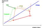
Brain perfusion pressure = BPav. - ICP = 75 - 85 mm Hg. Art.
Cerebral ischemia - with perfusion pressure< 50 мм рт. ст.
HAZARDS OF COMATOS
Violation of protective reflexes - aspirationViolations
breathing
–
violations
passability
respiratory tract, hypoventilation, atelectasis, apnea,
hyperventilation
Hemodynamic disorders
Hypo- and hyperthermia
Convulsive syndromes
Neuro-trophic disorders
Positional injury
lack of control of physiological items
Violations of energy and water-electrolyte balance
(dehydration, degeneration, immunodeficiency)
EXAMINATION OF THE PATIENT IN COMATIC CONDITION
1. Assessment of the respiratory and cardiovascular systems2. Neurological examination:
assessment of the level of consciousness and the depth of coma according to the Glasgow scale
examination of the eyes (uniformity and width of the pupils,
photoreaction)
assessment of muscle tone and reflexes
3. Clinical and laboratory examination:
general examination and objective examination
laboratory tests (blood sugar, blood gases, urea,
coma of unknown etiology - alcohol and other toxicological
analyzes, urine acetone, osmolarity, lactate, electrolytes,
leukocytosis, etc.)
instrumental
methodology
(lumbar
puncture,
echoencephaloscopy, CT, NMR - in case of suspicion of
intracranial pathology, EEG - in the diagnosis of death
brain)
LABORATORY DIAGNOSTICS OF COMA
CriteriaDiabetic
Blood sugar
mmol / l
Urine acetone
m.
Osmolarity
m.
blood pH
Hemoconcentration
Lactate
Hepatic
Uremic
Chlorpenic
Brain
Poisoning
m.
m.
m.
Bilirubin
Transaminases
m.
Ammonia
m.
Urea
m.
Chlorides
Potassium
Liquor
Toxicological
analyzes
m.
Erythrocytes,
leukocytes
+++
Differential diagnosis of coma in diabetes mellitus
KetoacidoticHyperosmolar
Lactic acidotic
Hypoglycemic
blood sugar,
mmol / l
14-30
33-70
N,
promoted
< 3,0
urine acetone
++++
-
-
-
osmolarity
<320
up to 450
N
N
lowered
(acidosis)
N
sharp
lowered
N
moderate
sharp
No
No
N
N
sharp
promoted
N
Criteria
pH of blood
hemoconcentration
lactate
PRINCIPLES OF COMATIC CONDITIONS THERAPY
1. Elimination of the cause of a coma;2. Ensuring adequate breathing:
protection and maintenance of the passability of the DP (installation
airway, if necessary, tracheal intubation);
Oxygen therapy;
normalization of sputum drainage;
respiratory support (with respiratory depression - mechanical ventilation);
3. Maintaining adequate blood circulation:
monitoring of hemodynamic parameters;
infusion therapy;
inotropic and vasopressor drugs;
Normalization of CODE (albumin); 4. Normalization of VEB and CBS indicators;
5. Regulation of adequate diuresis (impaired renal perfusion)
and functions of the gastrointestinal tract (intestinal paresis);
6. Normalization of microcirculation:
Anticoagulants and antiplatelet agents;
Dopamine (2-5 mcg / kg / min).
7. Normalization of the temperature balance;
8. Therapeutic (enteral and / or parenteral) nutrition;
9. Symptomatic therapy:
- measures to protect the brain (hypothermia,
sedation);
- correction of cerebral circulation;
- anticonvulsant therapy;
10. Intensive care - head position at an angle of 30-45
degrees, kinetic therapy, exercise therapy, oral hygiene,
skin, perineum, etc. COMA
DP
PASS
DP OBSTRUCTION
VENTILATION IS NOT DISTURBED
VENTILATION DISTURBED
HEMODYNAMICS IS NOT IMPAIRED
HEMODYNAMICS DISTURBED
IF HYPOGLYCEMIA IS SUSPECTED: 20-40 ml 40% glucose
FOR CONVILANCY SYNDROME: benzodiazepines, barbiturates
COMPLETE LABORATORY EXAMINATION OF THE PATIENT
OBJECTIVE INSPECTION OBJECTIVE INSPECTION
PHOTO REACTION (+)
REFLEXES ARE SYMMETRICAL
THERE ARE NO SIGNS OF TBI
PHOTO REACTION (-)
REFLEXES ARE ASYMMETRIC
THERE ARE SIGNS OF TBI
DIFFUSE LOSS
FOCAL DEFEAT
LUMBAL PUNCTION
LIQUOR ASSESSMENT
NORM
NEUROSURGICAL CONSULTATION
MUDDY, LEUKOCYTES HEMORRHAGIC
METABOLIC
COMA
SEEDING, ABT
COMPUTER
TOMOGRAPHY
SOLUTION OF THE QUESTION ABOUT
OPERATIONS
Severe TBI
Priority activitiesGive the patient a semi-sitting position with
raised head end of the bed by 25-45
degrees;
If the patient has signs of aspiration
syndrome and impaired consciousness (coma, deep
stupor) - immediate intubation is recommended
trachea and sanitation of TBD;
If there are solids in the aspirated liquid
food particles, the progression of acute respiratory
failure, an emergency medical diagnostic bronchoscopy is indicated.
Severe TBI
Attention. Early IVL and sedation are most effectivemeasures to prevent secondary brain damage.
Any of the following conditions is an indication for
IVL:
coma (3-8 points on the Glasgow scale);
hyper, - or hypoventilation syndrome;
violation of the rhythm of breathing;
signs of an increase in intracranial hypertension;
concomitant chest injuries;
traumatic shock;
signs of decompensated respiratory failure of any
genesis.
Orotracheal intubation is preferable
Synchronize with a ventilator using sedatives,
analgesics and non-depolarizing muscle relaxants;
No hypovolemia - 0.9% sodium chloride solution IV evenly in
during the day, 30-35 ml / kg / day. Mannitol (Mannitol) is administered in 15-20
minutes at the rate of 1 g / kg of body weight. Then - 3-4 times a day (0.5-1 g / kg) In cases of unclear diagnosis (coma of unexplained etiology)
patients should be referred to multidisciplinary
hospitals provided with round-the-clock duty
neurosurgeon, neuropathologist and therapist.
Transportation must be carried out with care, gentleness,
better by direct transport. If transplants
are inevitable, then it is necessary to transfer the patient to one and those
same stretcher. In the choice of transport and route
be guided by the patient's condition: choose the shortest
way, to spare the patient's head from jolts and abrupt changes in it
provisions. When using air transport, the height
flight should not exceed 3000 m. When using
road transport should be assessed and reminded
to the driver about a gentle ride. Before transportation
assess the state of the patient's vital functions and introduce
medications for stability during evacuation.
Slide 1
Slide 2
 * Definition of coma Coma - deep sleep (Greek) ... - a state of sharp inhibition of higher nervous activity, expressed by deep loss of consciousness and dysfunction of all analyzers - motor, skin, visual, auditory, olfactory and internal organs. (Bogolepov N.K. 1962)
Slide 3
 * Definition of coma ... - brain failure, expressed by a disorder of the self-regulating mechanisms of brain activity. (Kugelmass L.N. 1962) ... - an areactive state in which the awakening of the subject is impossible. (Plum F. and Posner L.B. 1966)
Slide 4
 * Stages of impairment of consciousness. Clear consciousness. Mild stunning - the ability to perceive speech with increased drowsiness (in the absence of aphasia). Deep stunning - the perception of simple speech with severe drowsiness. Stupor - executing only simple commands and opening your eyes to significant irritation. Moderate coma - lack of eyes opening and command execution, reactions to pain are differentiated. Deep coma - lack of opening the eyes and executing commands, reactions to pain - undifferentiated or posotonic. Atonic coma - atony, areflexia, normo- or hypothermia (it is possible to preserve spinal automatisms).
Slide 5
 * - The energy received by the brain is 95% -98% provided by glucose oxidation (without the participation of insulin) in the Embden-Meyerhof and Krebs cycles - Brain mass is about 2% of body weight - Volumetric blood flow is 20% of cardiac output
Slide 6
 Pathophysiological mechanisms of coma Any depression of the central nervous system (and even more so coma) has a biochemical or anatomical basis (or both at the same time)
Slide 7
 * Causes of coma Large-focal brain lesions: hematoma, brain abscess, brain tumor, epilepsy. Diffuse destructive lesions of brain tissue: contusion, encephalitis, meningitis, subarachnoid hemorrhage. Toxic brain damage: poisoning with alcohol and its surrogates, ethylene glycol, hydrocarbons and other poisons, mushroom poisoning, poisoning with drugs, sedatives, barbiturates, etc. Cerebral circulation failure: low emission syndrome, Edams-Stokes attacks, consequences of asystole, encephalopathy , ischemic stroke. Metabolic causes: disorders of water and electrolyte balance, hyperosmolar syndrome, hyper- or hyponatremia, acid-base disorders, calcium balance disorders, hypoxia, hyper- or hypocapnia, liver failure, uremia. Endocrine causes: hyper- or hypoglycemia, hyper- or hypothyroidism. Thermal homeostasis disorders: heatstroke, hypothermia
Slide 8
 * Most of the mechanisms of coma are associated with ischemic or traumatic destruction of neurons, oxygen deficiency in the brain tissues, and glucose uptake disorders.
Slide 9
 * Classifications of coma Scales of stages of coma development (stages, phases, levels) Scoring (point) systems
Slide 10
 * Scoring systems are based on the assessment of 3-4 (sometimes more) behavioral signs, for example: - the general orientation of the patient in the situation, time; - reactions to speech calls (verbal reactions); - physical activity; - reactions to painful irritations; - reactions of the cranial nerves; - the presence of seizures; - the nature of breathing;
Slide 11
 * Glasgow coma scale (Teasdale G. and Jennett B., 1974) Functional studies Score in points Spontaneous eye opening 4 to a speech command 3 to pain 2 no response 1 Motor response to a speech command 6 to pain irritation with pain localization 5 limb withdrawal with flexion 4 abnormal flexion of the limbs 3 extension according to the type of decerebral rigidity 2 no response 1 Speech reactions orientation and conversation 5 disorientation and conversation 4 incoherent words 3 incomprehensible sounds 2 no response 1 Oscillation limits: 3 - 15 points
Slide 12
 * Eye opening spontaneous 4 to speech command 3 to pain 2 no response 1
Slide 13
 * Motor response - to a speech command 6 - to painful irritation with localization of pain 5 - withdrawal of the limb with flexion 4 - abnormal flexion of the limbs 3 extension according to the type of decerebral rigidity 2 - no response 1
Slide 14
 * Verbal reactions - orientation and conversation 5 - disorientation and conversation 4 - incoherent words 3 - incomprehensible sounds 2 - no answer 1
Slide 15
 Absent (both pupils are fixed, cold test is negative) Present (at least on one side) Lack of response, apnea, collapse, polyuria, hypothermia 7 Outward coma Brain death Coma assessment scale (Bozza- Marubini M., 1983 ) Stimuli and responses Vocal Pain The most pronounced responses on the intact side Speech response to call 1 Mild drowsiness Execution of commands 2 Deep drowsiness Limited 3 Mild coma Withdrawal or abnormal flexion 4 Medium coma Nenor - minimal extension 5 Deep coma Assessment of stem reflexes by pupil response Abnormal flexion or extension or lack of response 6 Coma carus
Slide 16
 * Procedure for examining a patient in a coma Research of the functional state of the respiratory and circulatory systems to assess the viability of the body General clinical examination to assess extracranial pathology (supplemented by laboratory data). Elementary neurological examination to assess the functional state of the central nervous system and the depth of the coma.
Slide 17
 * Re-examination of the patient using the Glasgow and Glasgow-Pittsburgh scale is carried out no more than two hours later.
Slide 18
 * Cephalic reflexes characterizing coma (in descending order of diagnostic value) Pupillary Corneal Oculocephalic ("doll's eyes") Vestibular Audio-ocular (blinking) Proboscis Cough Pharyngeal Swallowing
Slide 19
 * The state of spinal reflexes (tendon, skin, muscle) in coma is not informative.
Slide 20
 * Differences in pupillary responses (dilation or constriction of the pupil on one side) and lateralization of other neurological symptoms are more indicative of the traumatic nature of coma.
Slide 21
 * Criteria for assessing breathing in coma Frequency Depth Rhythm Synchronism Criteria for assessing blood circulation in coma Heart rate Rhythm Blood pressure
Slide 22
 * Rough assessment of the key symptoms of the condition in coma Probability of an infectious process (sepsis, meningitis, pneumonia), heatstroke, damage to hypotomical structures in other forms of pathology. Hyperthermia
Slide 23
 * Rough assessment of the key symptoms of the condition in coma Probability of peripheral collapse, alcohol intoxication, barbituratomy poisoning, overdose of phenothiosin drugs, general hypothermia. Hypothermia
Slide 24
 * Rough estimate of the key symptoms of the condition in coma The probability of a volumetric process in the brain or intracranial hypertension of another origin. Hypertension
Slide 25
 * Rough estimate of the key symptoms of the condition in coma Probability of liver cirrhosis and hepatic coma. Edema and pronounced vascular pattern on the skin of the abdomen and chest
Slide 26
 * Rough assessment of the key symptoms of a coma condition The probability of cerebral hemorrhage, its tumor, diabetic coma, acidosis, uremia, ethylene glycol and methyl alcohol poisoning. Peripheral respiration of the Cheyne-Stokes and Kussmaul type
Slide 27
 * Rough estimate of the key symptoms of the condition in coma Probability of poisoning with drugs of the opium group or barbiturates. Constricted pupils with sufficient reaction to light.
Slide 28
 * Intracranial hypertension Intracranial hypertension in coma is always cerebral edema: Cytotoxic (swelling) Vasogenic
Slide 29
 * Therapy of cerebral edema: Maintaining adequate PaO2 (at least 60 mm Hg). Maintaining adequate cerebral blood flow (blood pressure avg more than 50 mm Hg) Maintaining normal plasma osmolality (285-295 mosmol / l). Steroid therapy. Diuretic therapy. Decompression of the cerebrospinal fluid space. Reduced water consumption. Hyperventilation. HBO. Barbiturate therapy.
Slide 30
 * Electroencephalography is not a specific diagnostic method, it has no signs of pathognomonicity. it is not very informative in assessing coma and especially in predicting its outcome. Methods of radiation diagnostics (Computed tomography, nuclear magnetic resonance, positron emission tomography, ultrasound - volumetric) Methods of choice for diff. diagnostics of the etiology of brain lesions
Slide 31
 * Clinical variants of coma Coma in traumatic brain injury Posthypoxic (postischemic) Coma in focal brain damage (stroke) Poisoning with drugs, barbiturates, psychotropic and sedative drugs Poisoning with alcohol and its surrogates Ketoacidotic Non-ketotic (hyperglycemic, hyperosmolar)
Slide 32
 * General principles of coma therapy Ensuring airway patency Monitoring and maintaining the optimal level of systemic circulation and blood oxygenation. Maintenance of water-electrolyte and protein balance of the body, periodic monitoring of electrolyte levels, plasma osmolality, biochemical parameters.
Slide 33
 * General principles of coma therapy Maintaining an optimal thermal balance. Sedation therapy with motor agitation (treatment with benzodiazepines), with convulsive syndrome, barbiturates are used. Forced ventilation of the lungs. Adequate therapy for cerebral edema
Slide 34
 * Features of diagnosis and treatment of coma in traumatic brain injury. It is impossible to delay the start of intensive treatment for the sake of a clarifying or even diagnostic neurological examination; Mandatory consultation with a neurosurgeon Mandatory puncture of the spinal canal; The detection of neurological asymmetry on examination is evidence in favor of a hematoma;
Slide 35
 * Features of diagnosis and treatment of coma in traumatic brain injury. It should be remembered that with hypotension, the results of neurological examination may be erroneous; Acidosis in patients with "traumatic" coma is always lactate and may require correction with bicarbonate; Treatment of cerebral edema is carried out only in the absence of signs of intracranial hematoma.
Slide 36
 * Diabetic coma Ketoacidotic coma. Hyperosmolar coma. Lactic acidosis coma. Hypoglycemic coma.
Slide 37
 * Reasons for decompensation Insufficient insulin administration Change of insulin preparation without determining the sensitivity to the new preparation. Violation of the injection technique. Unjustified termination of insulin therapy. Increased insulin requirements.
Slide 38
 * Decompensation of diabetes Manifestation of diabetic coma: Gastrointestinal form Cardiovascular form Encephalopathic form Renal form
Slide 39
 * Pathogenesis of ketoacidotic coma Toxic effect of excess ketone bodies> 3-5 mmol / L Hyperosmolarity Dehydration Hypoxia Acidosis
Acute liver failure (OPE). Definition, cause (etiology) of liver failure Kolomak V.A. ORENBURG REGIONAL MEDICAL COLLEGE
Acute liver failure (OPF) is a pathological syndrome based on acute damage to hepatocytes with subsequent disruption of their main functions (protein-educational, detoxification, production of blood coagulation factors, regulation of acid base balance, etc.) Kolomak V.A.

Kolomak V.A. 3

Kolomak VA.4 Classification I. In the course of the disease Acute Chronic II. In stages I, initial (compensated) II pronounced (decompensated) III terminal (dystrophic) ending in hepatic coma. IV hepatic coma

Kolomak V.A. 5 Clinical manifestations During the first stage Decreased and perverted appetite Weakness Nausea Decreased ability to work Aversion to food Emotional disorders During the second stage Jaundice Hemorrhagic diathesis Ascites Unmotivated weakness Dyspeptic disorders Hypoproteinemic edema During the third stage metabolic disorders Cachexia in other internal organs. Loss of consciousness. Spontaneous movements and reaction to pain at the onset of coma and subsequently disappear. Exotropia. Lack of pupillary reactions. Pathological (plantar) reflexes. Convulsions. Rigidity. EEG - slowing down the rhythm, decreasing the amplitude as the coma deepens.

Etiology. Acute liver failure (OPE) can occur against the background of the following diseases: 1. Causing damage to the hepatic parenchyma (acute and chronic hepatitis, cirrhosis, primary and metastatic liver tumors, echinococcosis, leptospirosis, yellow fever). 2. Complicated by cholestasis (choledocholithiasis, strictures of the biliary tract, tumors of the hepatic or common bile duct, pancreatic head, ligation or damage to the bile ducts during surgery, etc.). 3. Poisoning with hepatotropic poisons (chlorinated and aromatic hydrocarbons, chloroform, dichloroethane; ethyl alcohol, phenols, aldehydes, plant toxins, for example, pale toadstool) and drugs (drugs, chlorpromazine, etc.). 4. Diseases of the liver vessels (portal vein thrombosis). 5. Diseases of other organs and systems (endocrine, cardiovascular, infectious, diffuse connective tissue diseases). 6. Extreme effects on the body (trauma, burns, severe surgical interventions, prolonged compression syndrome) Kolomak V.А.

Forms of liver failure. Depending on the cause of the occurrence, there are endogenous, exogenous and mixed forms of OPF. At the heart of the endogenous hepatocellular form of acute hepatic failure of OPe is massive necrosis of the liver resulting from direct damage to its parenchyma. This condition can occur under the influence of the following factors: 1. Hepatotoxic and silver toxic effects of metabolites (tryptophan, tyrosine, methionine, butyric acid). 2. The appearance of false mediators replacing biogenic amines (norepinephrine, dopamine), which leads to disruption of the interaction of neurons. 3. Release and activation of lysosomal enzymes (especially hydrolases). 4. Cerebral edema with prolonged coma. 5. Violation of water-salt and acid base balance, leading to fluid retention in the extracellular space and a decrease in the BCC. 6. The emergence of coagulopathy (disseminated intravascular coagulation syndrome). 7. Accession of impaired renal function (ARF), lungs (distress syndrome). The exogenous (portocaval) form of acute hepatic failure, OPE, develops in patients with liver cirrhosis. Under normal conditions, 80% of endogenous ammonia is metabolized by the liver. With cirrhosis, this process is disrupted, resulting in damage to the central nervous system. The mixed form of acute liver failure OPEN usually occurs with a predominance of endogenous factors V.A. Kolomak.

Kolomak V.A.

Principles of treatment for acute renal failure: 1. Normalization of basic vital processes. 2. Conducting substitution therapy aimed at restoring lost or impaired body functions. Normalization of the main vital processes First of all, it is necessary to stop the necrosis of hepatocytes. For this purpose, the influence of hepatotoxic factors is eliminated or reduced: intoxication, bleeding, hypovolemia, hypoxia. 1. Normalization of basic vital processes. 1. Bleeding is stopped by a surgical or conservative method according to generally accepted indications and methods. 2. Hypovolemia is eliminated by introducing fluid into the body under the control of CVP and hourly diuresis. 3. Hypoxia is stopped by the normalization of lung function and hepatic blood flow. For this purpose, sorbitol (up to 1 g / kg), rheopolyglucin (up to 400 ml / day) are injected daily. An improvement in the blood supply to hepatocytes is achieved with xanthine drugs: euphyllin (10 ml 2.4% p-ra 34 times / day), sympatholytics (droperidol, pentamine), but without a sharp decrease in blood pressure.) Kolomak V.A.

4. Intoxication decreases with the elimination of intestinal paresis and cleaning it from protein half-life products, as well as restriction in protein intake or prescription of protein-free food. 5. The next stage of intensive therapy is to improve energy processes by introducing easily digestible ones. 6. The use of vitamins with hepatotropic action and various vitamins has a stimulating effect on the restoration of lost liver functions. 7. If solutions of amino acids are used in complex infusion therapy, they should not contain phenylalanine, tyrosine, tryptophan, methionine. Their introduction leads to increased disturbance of amino acid metabolism and the development of encephalopathy. 8. To reduce the flow of toxic products from the intestine to the liver, the intestinal microflora is suppressed. 9. In case of severe violations of the urea educational function of the liver, it is necessary to inject a solution of arginine chloride by intravenous drip at the rate of mg / kg / day. Normalization of basic vital processes (continuation) Kolomak V.A.

Substitution therapy for acute liver failure (ARF). 1. Hypoproteinia is arrested using albumin 2. For the correction of hemorrhagic syndrome, vikasol is used, fresh frozen plasma, aminocaproic acid 3. The detoxification function is interfered with the use of hemodesis (At the initial stages of the disease, hemo- and lymphosorption is indicated. 4. Correction of water-electrolyte metabolism and acid base balance. 5. С the initial stages of treatment are preventive treatment Kolomak V.A. homo-liver, liver transplantation, exchange blood transfusion, cross circulation, etc. Kolomak V.A.